|
|
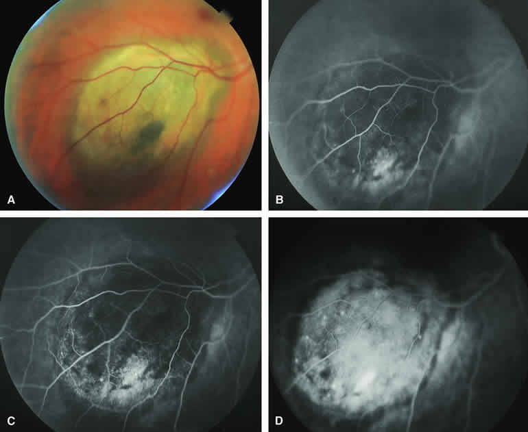
| Fig. 9. Typical melanotic choroidal melanoma without invasive features. A. Nodular gray-brown nodular choroidal tumor with linear clumps of orange lipofuscin pigment and central whitish discoloration of overlying retinal pigment epithelium. B-D. Fluorescein angiogram of lesion. B. Venous phase frame showing mild hypofluorescence of most of tumor but with ill-defined hyperfluorescent focus near inferior margin. C. Later venous phase frame showing persistent generalized hypofluorescence of mass, increased smudgy and punctate hyperfluorescence near the inferior and temporal margins of lesion, and choroidal fluorescence blockage by lipofuscin pigment overlying mass. D. Late-phase frame showing diffuse hyperfluorescence of subretinal fluid and outer retina overlying mass, persistent hypofluorescence corresponding to the margin of the lesion, persistent choroidal fluorescence blockage corresponding to the lipofuscin pigment clumps, and several pinpoint dots of intense hyperfluorescence overlying the mass. |