Chapter 113F
The Use of Fluorescein Angiography in Acquired Macular Diseases
ANTONIO P. CIARDELLA, STEPHEN R. KAUFMAN and LAWRENCE A. YANNUZZI
Main Menu   Table Of Contents

Search

AGE-RELATED MACULAR DEGENERATION
CENTRAL SEROUS CHORIORETINOPATHY
EPIRETINAL MEMBRANE
MACULAR CYSTS, HOLES, AND PSEUDOHOLES
CYSTOID MACULAR EDEMA
MYOPIC MACULOPATHY
TRAUMA-RELATED MACULOPATHY
MISCELLANEOUS CONDITIONS
PHOTIC MACULOPATHY
CONCLUSIONS
REFERENCES

Fluorescein angiography (FA) has been widely used clinically for more than 3 decades, and it has been valuable in the understanding, diagnosis, and treatment of acquired macular diseases. Extensive use of FA, combined with growing knowledge of the range of clinical presentations and natural histories of the acquired macular diseases, has helped clinicians obtain an appreciation of the indications for FA. Our aim in this chapter is to illustrate useful parameters of FA and to provide guidance for the optimal use of this technique. Comprehensive reviews of the interpretation of the fluorescein angiogram may be found elsewhere.1,2
Back to Top
AGE-RELATED MACULAR DEGENERATION

DIAGNOSIS

Age-related macular degeneration (AMD) may be divided into two types. Nonexudative (“dry”) AMD has several morphologic forms, including “hard” discrete drusen, shallow retinal pigment epithelial detachments associated with thickened Bruch's membrane (“soft” drusen), and geographic atrophy (GA) of the retinal pigment epithelium (RPE).3 On FA the area of GA appears hyperfluorescent for window defect from the early frames of the angiogram, with late staining of the underlying sclera (Fig. 1). However, these pathologic changes can usually be assessed by clinical examination, and FA is generally not necessary to diagnose nonexudative AMD. An exception is cuticular drusen, which may appear clinically as a subtle disturbance of the RPE; FA reveals multitudes of small, discrete drusen described as “stars in the sky” (Fig. 2). The second type of AMD, which is associated with soft drusen, is known as exudative (“wet”) AMD. It is due to a choroidal neovascular membrane that has incompetent vessels resulting in detachments of the RPE and the neurosensory retina. Consequently, in patients with a large RPE and/or serous neurosensory detachment, FA is often necessary to rule out a choroidal neovascularization (CNV). In general, a small pigment epithelium detachment (PED) and a larger neurosensory detachment overlie CNV, while the opposite is generally the case in a nonexudative PED. Additionally, CNV often presents as a “notched” PED (Fig. 3).4 The presence of subretinal blood or pigment at the border of a PED strongly indicates that the detachment is exudative in origin (Fig. 4). Similarly, a rip in the RPE generally reflects subretinal fibrosis from a CNV (Fig. 5 and 6). The diagnosis is more difficult in patients who have a chronic, organized PED. Such a lesion may be due to either nonexudative AMD or to an organized, fibrotic CNV. Clinically and angiographically, it may be impossible to distinguish between these two conditions. In most cases, however, FA does assist in making the diagnosis.

Fig. 1. Late-phase fluorescein angiography of an eye with a central area of geographic atrophy, which appears hyperfluorescent. A few soft drusen are also present.

Fig. 2. Multiple cuticular drusen and subretinal neovascularization. This patient had some central soft drusen, which, unlike cuticular drusen, are associated with neovascularization. The inferior aspect of the choroidal neovascular membrane has blocked fluorescence due to hemorrhage. Note the presence of the small, hyperfluorescent cuticular drusen that confer a “stars in the sky” appearance.

Fig. 3. Notched retinal pigment epithelium (RPE) detachment. There is a notch at the superotemporal border of a large RPE detachment that fills unevenly with fluorescein (large arrowhead). There is also a superior neovascular complex (small arrowhead) that hyperfluoresces. (Courtesy of Dr. Kenneth G. Noble.)

Fig. 4. Subretinal blood from a choroidal neovascular membrane. A small hemorrhage has layered out at the inferior aspect of a large retinal pigment epithelium (RPE) detachment. A shallow overlying neurosensory detachment can be appreciated as the slightly darkened, narrow band that surrounds the RPE detachment. The neurosensory detachment is being filled with fluorescein through a break in the RPE. The subretinal neovascular membrane, not clearly evident in this early-phase fluorescein angiogram, is at the nasal edge of the RPE detachment, near the optic disc.

Fig. 5. A. Clinical photograph of a large, crescent-shaped rip of the retinal pigment epithelium (RPE) in the temporal macula. B. Early-phase fluorescein angiography study demonstrates the presence of a window defect corresponding to the RPE rip, which exposes the choroidal vasculature. Where the RPE is redundant in the central macula there is blockage of the normal choriocapillaris fluorescence. C. Late-phase angiogram reveals intense hyperfluorescence seen through the RPE defect.

Fig. 6. Classic, submacular choroidal neovascularization (CNV) and retinal pigment epithelium (RPE) rip. Fluorescein angiography reveals the presence of a well-defined subfoveal CNV and hyperfluorescent area in the inferior macula corresponding to the RPE rip (yellow arrow).

In patients with a shallow neurosensory detachment, the Amsler grid test and visual acuity may be normal. If there is subtle elevation of the neurosensory retina on biomicroscopy examination, FA may demonstrate a CNV before it is symptomatic. It is often easier to evaluate both RPE and neurosensory detachments with good stereoscopic FA pictures than with direct examination.5 Consequently, FA can be helpful in determining the presence and extent of these processes. This is particularly important in patients with CNV due to AMD, because its aggressive course often requires prompt intervention to save central vision.6,7

Furthermore, FA helps in recognizing two types of CNV: classic and occult. Classic CNV consists of a well-defined neovascular membrane, which is apparent in the early phase of the angiogram and shows late leakage of dye beyond its boundaries (Fig. 7 and 8). Occult CNV is seen on by FA as an area of late hyperfluorescence of undefined origin or as a neovascularized PED (Fig. 9 and 10 ). Mixed-type CNV is predominately classic or minimally classic depending on whether the classic component is more or less than 50% of the entire lesion (Fig. 11).

Fig. 7. Composite photograph of fluorescein angiography study in a patient with classic, subfoveal choroidal neovascularization (CNV) in the right eye. A The classic neovascular membrane appears as a well-defined area of hyperfluorescence in the early phases of the angiogram. There is leakage of dye from the classic net in the subretinal space throughout the study. B. In the late phase of the study, the edges of the CNV are fuzzy and indistinguishable.

Fig. 8. A. Color photograph of subfoveal classic choroidal neovascularization (CNV). The neovascular membrane appears as a dirty gray, subfoveal lesion surrounded by exudative neurosensory detachment. B–D. Fluorescein angiography demonstrates early hyperfluorescence and late leakage of the CNV.

Fig. 9. A. Clinical photograph of the left eye of a patient with exudative neurosensory macular detachment. There were also intraretinal and subretinal hard exudates, subretinal hemorrhage, and retinal pigment epithelium (RPE) changes. B–D. Fluorescein angiography of the same eye demonstrates the presence of stippled hyperfluorescence from the RPE, and late-phase oozing of dye of undefined origin. There was occult choroidal neovascularization.

Fig. 10. A. Red-free photograph of the right eye of a patient with wet age-related macular degeneration. There was a large, exudative pigment epithelium detachment (PED), with a narrow band of subretinal hemorrhage at its inferior border. A notch in the PED is present at its nasal edge. There were also soft drusen. B–C. Fluorescein angiography demonstrates pooling of dye into the PED (short arrows). There was also late hyperfluorescence of undefined origin consistent with occult choroidal neovascularization (long arrows). There was blockage of fluorescence at the inferior border of the PED caused by subretinal hemorrhage.

Fig. 11. A. Red-free photograph of the right eye of a patient with wet age-related macular degeneration reveals exudative, neurosensory detachment in the macula and a few subretinal hemorrhages. B. Early-phase fluorescein angiography demonstrates well-defined classic choroidal neovascularization (CNV) (arrowhead). C. Late-phase fluorescein angiography shows leakage of dye from the classic CNV surrounded by an area of late hyperfluorescence consistent with occult CNV (arrows).

FA is also useful in characterizing two other subgroups of CNV: retinal angiomatous proliferation (RAP)8–16 and polypoidal choroidal vasculopathy (PCV).17–50 RAP begins in the deep retinal complex, forming intraretinal neovascularization (IRN), which may subsequently progress to extend beneath the neurosensory retina, forming subretinal neovascularization (SRN), and a vascularized PED.8 In the later phases of the process there may be a retinal-choroidal anastomosis (RCA). Clinical features of RAP include intraretinal hemorrhages, cystoid macular edema, and associated vascularized PED. FA is useful in revealing the presence of the angiomatous intraretinal vascular complex and the extension of the associated PED (Figs. 12 and 13). However, other diagnostic techniques such as indocyanine green (ICG) angiography, and optical coherence tomography (OCT) may be able to better demonstrate the presence of the RAP lesion.

Fig. 12. A. Clinical photograph of a retinal angiomatous proliferation (RAP) lesion (arrow). Note the intraretinal angiomatous proliferation, a feeding retinal arteriole, and a draining retinal venule, as well as the presence of intraretinal hemorrhages. B–C. Fluorescein angiography reveals late leakage from the RAP lesion.

Fig. 13. A. Early-phase Fluorescein angiography demonstrating the presence of an intraretinal angiomatous lesion (arrow). There is an associated pigment epithelium defect (PED), which is still hypofluorescent. B. Late-phase fluorescein angiography shows leakage from the retinal angiomatous proliferation (RAP) lesion and polling of dye into the PED. C. Indocyanine green angiogram of the same eye better demonstrates the presence of a hot spot corresponding to the RAP lesion. The PED remains hypofluorescent. D.Optical clearance tomography image demonstrates the presence of a serous PED and of intraretinal neovascularization.

PCV is characterized by the presence of dilated, choroidal vascular channels ending in orange bulging polyp-like dilations in the peripapillary and macular area. Associated features are recurrent subretinal hemorrhage and vitreous hemorrhage, relatively minimal fibrous scarring, absence of retinal vascular disease, pathologic myopia, and signs of intraocular inflammation. FA demonstrates the presence of the dilated vascular channel (Fig. 14 and 15). However, the presence of blood and exudation may block the details of the choroidal circulation on the angiogram. In these cases, ICG angiography can better demonstrate the presence of a distinct network of vessels within the choroid because the larger choroidal vessels are filled with dye.

Fig. 14. A. Color photograph of the right eye shows a ramified pattern of choroidal vascular abnormality irradiating from the peripapillary area toward the macula. The dilated vascular channels end with bulging polyp-like structures. A larger, orange, saccular dilation is seen inferior to the macula (white arrow); leakage of fluid from this vascular abnormality results in serosanguineous pigment epithelium detachment (black arrows). B. The corresponding fluorescein angiogram composite highlights the vascular lesion in the peripapillary area and the serosanguineous detachment of the pigment epithelium that extends inferiorly and temporally off the macula.

Fig. 15. A 51-year-old Caucasian woman was referred with diagnosis of central serous chorioretinopathy in her right eye. A and B. Color photograph and red-free photograph of the right eye show the presence of atrophic changes in the retinal pigment epithelium consistent with central serous chorioretinopathy. C, Fluorescein angiogram of the right eye reveals the presence of avascular network of small-caliber vessels terminating in polyp-like structures. D. Indocyanine green angiogram of the right eye confirms the presence of the polypoidal lesion.

The FA can also distinguish CNV from simulating lesions. For example, a dark mound of blood due to hemorrhage from a CNV will block choroidal fluorescence, whereas vascular tumefactions such as choroidal hemangiomas leak fluorescein. Choroidal melanomas frequently block early choroidal fluorescence and then leak fluorescein from their intrinsic vascular network in later phases of FA.

TREATMENT

FA is vital for the management of CNV.51–55 It can define the borders of the membrane and help localize the fovea. The final determination whether the CNV is subfoveal, juxtafoveal, or extrafoveal requires use of both FA to outline the membrane with respect to the retinal vasculature and clinical examination to define the precise location of the fovea (Fig. 16). Some patients, particularly those with high myopia, have an indistinct foveal avascular zone on FA. Other FA clues, such as the location of the macula lutea pigment, can be deceptive, because fixation does not necessarily correspond to the center of the macula lutea. The value of FA is also limited in cases of occult or poorly defined CNV, in which the exact location of the leaking CNV vessels cannot be angiographically determined (Fig. 17), and in patients with subretinal hemorrhage that obscures the membrane. In these patients, ICG angiography may be the most precise means of localization.

Fig. 16. Subfoveal, juxtafoveal, and extrafoveal choroidal neovascular membranes. A and B. Large subfoveal choroidal neovascularization (CNV) in a 69-year-old man with blood and pigment blocking central fluorescence on both the early-phase (A) and late-phase (B) photographs. The hypofluorescence surrounding the membrane is commonly seen in CNV and may be due to lipofuscin. C. Juxtafoveal CNV in a 37-year-old man with idiopathic CNVM. D. Cuticular drusen in same patient as in C were asymptomatic. E. Years later, this same patient developed a large extrafoveal CNV with central macular pigment abnormalities. A large neurosensory detachment was responsible for the disappearance of the drusen. (Courtesy of Dr. Kenneth G. Noble.)

Fig. 17. Poorly defined choroidal neovascularization (CNV). This woman has a CNV that hyperfluoresces in the juxtapapillary area; however, its full extent cannot be determined owing to blockage of fluorescence by blood. (Courtesy of Dr. Kenneth G. Noble.)

Conventional laser thermophotocoagulation is the treatment of choice for extrafoveal, well-defined, classic CNV. Photodynamic treatment (PDT) is the treatment of choice for subfoveal, predominantly classic CNV. FA is used to localize the lesion in relation to the fovea, classify the subtype, choose the type of procedure, and guide the treatment (Figs. 18, 19, and 20).56–73

Fig. 18. A. Late-phase fluorescein angiogram demonstrates the presence of actively leaking, subfoveal, classic choroidal neovascularization. B. After photodynamic treatment (PDT) with Visudyne there was complete closure of the neovascular membrane. The round, hypofluorescence corresponds to the area treated with PDT. There was no damage to the retinal vasculature.

Fig. 19. A. Red-free photograph of the right eye of a patient with exudative angiomatous macular degeneration. B. Fluorescein angiography reveals the presence of subfoveal, classic choroidal neovascularization (CNV). The boundaries of the CNV are digitally traced (yellow), and the greater linear dimension of the lesion is measured (red) to guide the PDT.

Fig. 20. A. Red-free photograph of a 20-year-old patient with sudden loss of vision to the level of 20/200. There is exudative, neurosensory macular detachment, a few hemorrhages, and lipid exudates. B. Fluorescein angiography reveals the presence of classic choroidal neovascularization (CNV), which appears to be juxtafoveal (<200 μ from fixation). Given the size of the CNV and its proximity to the fovea, it was decided to treat the patient with photodynamic treatment (PDT). C. Red-free photograph of the same eye 2 weeks after PDT; there is increased subretinal exudation D. Fluorescein angiography demonstrates that the CNV is still actively leaking. E. Red-free photograph 4 weeks after PDT demonstrates further increase in the size of the neurosensory macular detachment, subretinal hemorrhages, and lipid exudation. F. Fluorescin angiography reveals that the CNV has extended under the fovea. Given the young age of the patient, an inflammatory component of the neovascular process was suspected. It was decided to give a posterior, subtenon injection of triamcinolone acetonide, 40 mg/1 mL. G. Two weeks after steroid treatment there is partial reabsorption of the subretinal fluid. H. Fluorescein angiography demonstrates contraction of the CNV. I. Four weeks after injection of triamcinolone there is further reduction in the degree of neurosensory detachment; vision had improved to 20/60. J. Fluorescein angiography demonstrates that the CNV is smaller and less active (less leakage).

FOLLOW-UP

FA is needed to assess response to laser photocoagulation of a CNV and to diagnose recurrent membranes.51,54 The authors generally obtain angiograms 2 weeks, 1 month, 3 months, and 6 months after treatment. The risk of recurrence is greatest during the first 3 months, and the patient, who often has decreased vision due to prior neurosensory detachment, may be asymptomatic. FA is also needed to evaluate the results of PDT. In the original protocol of the Verteporfin in Photodynamic Therapy (VIP) and Treatment of Age-Related Macular Degeneration with Photodynamic Therapy (TAP) studies, a fluorescein angiogram was obtained every 3 months, and if there was persistent leakage from the CNV PDT was applied again (see Fig. 1820).60

Back to Top
CENTRAL SEROUS CHORIORETINOPATHY

DIAGNOSIS

Central serous chorioretinopathy (CSC) is characterized by breakdown of the outer retinal barrier, with leakage of fluid through a defect in the retinal pigment epithelium into the subretinal space, resulting in a serous neurosensory detachment.78–205 The ophthalmologist can usually diagnose CSC based on the clinical examination and demographic information.93–95 Most patients with CSC are middle-aged men74 who often have type A personalities.75, 96–104 CSC has also been associated to the use of corticosteroids,105–118 pregnancy,119–126 increased adrenaline level and stress,127–132 hemodialysis,133,134 collagen vascular diseases,135–147 and hypertension.148–157 CSC typically presents as a large serous detachment in the posterior pole without an obvious source of the subretinal fluid.76 However, because a small CNV cannot be ruled out, FA is usually done to confirm the diagnosis. Characteristically, there is a small RPE defect, which hyperfluoresces early, and then there is slow filling of the overlying neurosensory detachment, which may have a classic “smokestack” (Fig. 21) or “ink blot” (Fig. 22) appearance.158–161 Occasionally, FA demonstrates multiple sites of leakage (Figs. 23, 24, and 25). FA sometimes fails to distinguish CSC from CNV readily because fibrinous subretinal precipitates can cause slow filling of the RPE detachment, which is suggestive of CNV (Fig. 26). Sometimes peripapillary PCV can cause a neurosensory macular detachment masquerading as CSC (Fig. 27).77

Fig. 21. Central serous chorioretinopathy. A. There is a central retinal pigment epithelium (RPE) detachment with linear RPE hyperplasia in a “hot-cross bun” configuration, indicating that the RPE detachment is chronic. B. This late fluorescein photograph illustrates leakage of fluorescein into the neurosensory detachment, with a classic “smokestack” formation.

Fig. 22. Central serous chorioretinopathy. A. In this arterial-phase photograph, a small area of early hyperfluorescence represents a small RPE detachment in the inferotemporal macula. B. This mid-phase photograph reveals increased fluorescence in the area, reflecting filling of a neurosensory detachment. C. The detachment continues to fill with fluorescence in this late-phase photograph. This gradually enlarging fluorescence is sometimes called “ink blot” filling of the subretinal space. (Courtesy of Dr. Kenneth G. Noble.)

Fig. 23. Central serous chorioretinopathy with multiple retinal pigment epithelium (RPE) detachments. A. This red-free photograph demonstrates the large macular neurosensory detachment. B. Early-phase fluorescein angiogram reveals three hyperfluorescent RPE defects. C. These defects do not contribute evenly to the neurosensory detachment. Although the superior macular defect is leaking fluorescein into the subretinal space intensely, the defect near the optic nerve head along the superotemporal arcade is not leaking fluorescein and does not have an overlying neurosensory detachment. D. After treatment of the RPE detachments, the neurosensory detachment has resolved. (Courtesy of Dr. Kenneth G. Noble.)

Fig. 24. A 34-year-old Caucasian man presented, complaining of blurred vision in his left eye of 1-week duration. A. Clinical photograph of the left eye shows serous neurosensory macular detachment. B and C. Fluorescein angiography demonstrates a pinpoint area of hyperfluorescence in the central macula, leading to the characteristic smokestack configuration seen in the late-phase angiogram. A pigment epithelial detachment temporal to the fovea is increasing in hyperfluorescence throughout the study (white arrows).

Fig. 25. A 41-year-old man with the diagnosis of central serous chorioretinopathy in both eyes for 10 years presented complaining of sudden decrease of vision in the left eye. A. Red-free photograph of the right eye reveals pigmentary changes temporal to the fovea. B. Red-free photograph of the left eye shows a well-circumscribed neurosensory detachment of the macula with two areas of focal pigment epithelium detachment (PED). C– F. Fluorescein angiography shows two localized areas of PED and the typical “smokestack” appearance of the dye leaking under the detached retina. Note that the dye expands in an umbrella-like fashion once it reaches the upper limit of the detachment. G. Mid-phase fluorescein angiography of the fellow eye shows window-defect hyperfluorescence corresponding to the retinal pigment epithelium tract that extends inferiorly.

Fig. 26. A 36-year-old man presented with decreased visual acuity in the right eye. A. Color photograph of the right eye shows serous neurosensory detachment (white arrows) in the superior area of the macula with ring-like yellowish subretinal nodular deposits consistent with fibrin surrounding the localized pigment epithelium detachment (PED) (black arrows). B. Early-phase fluorescein angiography reveals a localized area of leakage corresponding to serous PED. C. Late-phase fluorescein angiography demonstrates hyperfluorescence due to pooling beneath the serous PED. D. Late-phase indocyanine green angiogram shows an area of hyperfluorescence corresponding to the serous PED with staining of the subretinal fibrin deposits.

Fig. 27. A. Clinical photograph of a 62-year-old woman with a neurosensory retinal detachment in the central macula. B. Indocyanine green angiogram reveals the presence of a polypoidal choroidal vascular abnormality in the superior temporal juxtapapillary region.

The diagnosis of CSC may be difficult if there is neither RPE detachment nor evidence of leakage into the subretinal space. Possible causes of a neurosensory elevation without evidence of leakage in the macula include CSC with the RPE detachment located outside the macular area (Fig. 28), CSC with a healed leak (in which case the neurosensory detachment should resolve soon), peripheral retinal hole, choroidal tumor, congenital optic nerve pit, and idiopathic uveal effusion syndrome. There are several other considerations in a patient who presents with a localized serous detachment of the macula, including age-related macular degeneration,162–164 a macular hole in a patient with high myopia, malignant hypertension, toxemia of pregnancy, collagen vascular disease, disseminated intravascular coagulation, choroidal inflammatory disease, Coat's disease (Fig. 29), and ocular contusion. Usually, these conditions are diagnosed based on clinical examination. ICG angiography may be helpful in differentiating CSC from AMD. On ICG studies there is often diffuse choroidal hyperpermeability in patients with CSC.165–181

Fig. 28. A 30-year-old woman had loss of vision in the right eye to 20/80. The patient was on systemic corticosteroid treatment for renal transplant rejection. A. Clinical photograph of the right eye demonstrates a neurosensory macular detachment. B. Late-phase fluorescein angiography reveals the presence of three3 actively leaking PEDs above the optic disc. Note the similarity between this case and the case illustrated in Figure 27 with PCV.

Fig. 29. 14-year-old adolescent girl with sudden loss of vision in the left eye to 20/400. A. Clinical photograph of the posterior pole demonstrates a neurosensory macular detachment simulating central serous chorioretinopathy. B. Composite fundus photograph of the same eye demonstrates the presence of a localized area of retinal capillary telangiectasia in the temporal periphery. The intraretinal yellowish material is consistent with dehemoglobinized blood. There are also intraretinal exudates scattered throughout the fundus. C. Composite fluorescein angiogram of the same eye reveals diffuse retinal capillary telangiectasia and intraretinal leakage. The dilated retinal telangiectatic vessels are actively leaking and are responsible for the macular neurosensory detachment. This patient was diagnosed with Coats' disease. D. Scatter laser photocoagulation of the telangiectatic vessels was carried out.

TREATMENT

The clinical course of CSC tends to be benign, with complete resolution within 3 to 4 months.74 Focal laser treatment of small RPE detachments has never been shown to improve the long-term vision of patients with CSC,78 but it does hasten resolution of the neurosensory detachment and it reduces the recurrence rate.79,182–203 The best candidates for treatment include those who have occupational needs and are strongly motivated to undergo treatment, and those with a chronic neurosensory detachment of 4 months or longer, particularly if there is evidence of loss of central or paracentral vision.55 Laser treatment to the point of RPE leakage is very effective in resolving the neurosensory detachment.80,81 In chronic CSC, grid treatment reduces macular edema and lipid and tends to stabilize vision.82,83

FOLLOW-UP

FA is rarely needed to follow the course of patients with CSC. As the subretinal fluid resolves, patients will have improved visual acuity, decreased metamorphopsia, and decreased induced hyperopia. Contact lens biomicroscopy reliably evaluates the amount of subretinal fluid. If the neurosensory detachment persists for several weeks after laser treatment, FA can be valuable in elucidating whether the RPE detachment has failed to resolve despite treatment, whether there have been unnoticed RPE defects that were not treated, whether there is recurrent CSC, or whether there is another cause of neurosensory detachment.

Although several authorities consider CSC to be a relatively benign disease,84 some patients develop recurrent, chronic neurosensory detachments that slowly cause photoreceptor degeneration. These patients often have a diffuse RPE “ooze,” reflecting generalized RPE dysfunction (Fig. 30). Their persistent neurosensory detachment can eventually lead to significant visual loss.85

Fig. 30. Diffuse retinal pigment epitheliopathy variant of central serous chorioretinopathy (CSC). Repeated episodes of acute CSC can result in chronic retinal pigment epithelium dysfunction with “ooze” of fluorescein. Unlike acute CSC, this uncommon variant often causes substantial loss of central vision.

Clinically discernible peripheral dependent bullous neurosensory detachments have been described in patients with chronic CSC.85–92 Yannuzzi and co-workers first characterized the presence of RPE atrophic tracts extending inferiorly in the fundus periphery secondary to antecedent retinal detachment in patients with CSC.85 Presumably, there is a particularly severe and/or longstanding leakage of fluid from an RPE defect in the subretinal space at the posterior pole. The subretinal fluid gravitates inferiorly to form a dependent neurosensory detachment in a “flask,” “teardrop,” “dumbbell,” or “hourglass” pattern (Fig. 31). Sometimes the tract of subretinal fluid connecting the macular detachment with the bullous neurosensory detachment in the inferior hemisphere is so shallow that it is very difficult to appreciate. The RPE under the chronic retinal detachment experiences atrophic changes that appear as atrophic RPE tracts connecting the posterior pole with the dependent retinal detachment. The retina itself develops secondary manifestations including pigment migration, capillary dilatation (telangiectasia) proximally and capillary nonperfusion (ischemia) distally to the area of detached retina (see Fig. 31). The changes in the RPE consist of both RPE atrophy and pigment clumping in the form of perivascular deposits or bone spicules, a condition described by Gass as a “pseudoretinitis pigmentosa–like atypical CSC presentation.”87

Fig. 31. A 47-year-old woman with an18-year history of central serous chorioretinopathy in both eyes. A. Color photograph composite of the left eye shows bullous dependant detachment of the neurosensory retina inferiorly. B. Fluorescein angiogram composite reveals diffuse decompensation of the retinal pigment epithelium, multiple scattered pigment epithelium detachments 9PEDs), and obliteration of the retinal capillaries in the region of the detachments. Note the presence of early neovascularization at the junction between perfused and non-perfused retina. C. Clinical photograph of the left eye shows PED superior to the optic disc partially surrounded by fibrin deposits. D. Fluorescein angiography confirms the presence of active leakage from the serous PED. E, Color photograph composite of the same eye 2 months after laser treatment of the site of leakage reveals partial resolution of the detachment and lipid precipitation. F. Clinical photograph composite 16 months after the laser treatment in the area of the leakage shows complete resolution of the detachment and partial reperfusion of the inferior retina.

Other complications noted in these patients are cystoid macular edema or, more frequently, cystoid retinal changes in the areas of chronic detachment, subretinal lipid deposition, choriocapillaris atrophy secondary to the RPE damage in the areas of RPE tracts, and CNV.89,90,204–206 This severe variant of CSC appears to be more frequent in patients of Latin or Asian ancestry, and it is usually associated with frequent recurrences, permanent central vision loss, and significant superior visual field loss.

Back to Top
EPIRETINAL MEMBRANE

DIAGNOSIS

Epiretinal membrane (ERM) formation has been associated with retinal vascular occlusive disease, diabetes, uveitis, penetrating or blunt trauma with intraocular hemorrhage, ocular inflammation, ocular surgery, laser or cryotherapy for retinal breaks, telangiectasia, macular holes, retinal angiomas, retinal arteriolar macroaneurysms, intraocular tumors, and retinitis pigmentosa.207 A preretinal sheen, readily appreciated on direct ophthalmoscopy or contact lens biomicroscopy, provides a diagnosis. In more severe cases, a fibrous membrane and distortion of the retinal vessels can be seen (Fig. 32). Consequently, FA is generally not necessary to confirm the diagnosis of ERM. Nevertheless, FA can demonstrate leakage from vessels that have become relatively incompetent owing to traction from the membrane (Fig. 33).208,209 This often accounts, in part, for loss of vision in symptomatic patients. OCT examination is the gold standard for the study of the vitreoretinal interface. It nt only demonstrates the presence of an ERM,, but also helps in quantifying the degree of tractional neurosensory elevation and in differentiating an ERM from vitreomacular traction syndrome.

Fig. 32. Preretinal gliosis. A. Red-free photograph demonstrates stretching of retinal vessels in the inferior macula toward the inferior arcade. The preretinal membrane apparently derives from a dense condensation of fibrous tissue in the inferior macula. The superior half of the macula is partly obscured by a fibrous condensation from posterior vitreous detachment. B. Fluorescein angiogram also demonstrates the traction on the retinal vessels, and the dense gliotic membrane partially blocks background fluorescence. There is some mild telangiectasia of the perifoveal retinal capillaries.

Fig. 33. Cystoid macular edema due to preretinal gliosis. A. Preretinal gliotic membrane has stretched retinal vessels, particularly nasally, toward the central macula. There is telangiectasia of the perifoveal capillaries and some early leakage superotemporally. B. Lat-phase photograph reveals cystoid accumulation of fluorescein. (Courtesy of Dr. Kenneth G. Noble.)

TREATMENT

An ERM that causes severe loss of vision may warrant vitrectomy and membrane peeling. In these cases, the membrane is clinically obvious and FA is not necessary. Ultrasound is often useful to assess the degree of posterior vitreous separation from the posterior pole.210–212

FOLLOW-UP

FA plays no role in the chronic management of patients with ERMs. The clinical presentation, including visual acuity, Amsler grid, and fundus appearance, guides long-term care.

Back to Top
MACULAR CYSTS, HOLES, AND PSEUDOHOLES

DIAGNOSIS

In general, the diagnosis of a macular hole is readily made in patients who present clinically with a full-thickness retinal defect, yellow exudative deposits at the base, and, occasionally, an operculum. However, it is sometimes difficult, using contact lens biomicroscopy alone, to distinguish a full-thickness macular hole from simulating conditions. Visual acuity is often 20/200 to 20/400 in an eye with a full-thickness hole, whereas eyes with macular cysts or pseudoholes usually have better vision. Visual acuity, however, cannot rule in or rule out a full-thickness macular hole.213,214

The differentiation of a partial-thickness, or lamellar, macular hole from a full-thickness macular hole can be challenging, and FA is often helpful in assisting in the diagnosis.55 As in a full-thickness hole, a lamellar hole presents as an excavation of the retina, and the presence of drusen underneath the lamellar hole can simulate the yellow deposits seen at the base of full-thickness holes. Occasionally, there is a full-thickness hole at one side of a lesion, and the rest of the lesion consists of a lamellar defect. FA will show immediate hyperfluorescence from the choroidal circulation under a full-thickness hole (Fig. 34A), whereas a lamellar hole, with a relatively intact RPE, will block some of the normal fluorescence from the choriocapillaris (Fig. 34B).

Fig. 34. Full-thickness and partial macular holes. A. There is a discrete hole in the central macula. Hyperfluorescence reflects choriocapillaris leakage that is normally partially blocked by retinal pigment epithelium. B. There is partial blockage of the choroidal hyperfluorescence, but subtle hyperfluorescence is visible. (Courtesy of Dr. Peter Judson.)

There are several causes of pseudoholes, including an area of sharply demarcated RPE atrophy, a dilated perifoveal capillary net, and an ERM with a clear center. Careful contact lens biomicroscopic examination will usually distinguish these conditions from a full-thickness macular hole, but FA can help. Both RPE atrophy and a full-thickness hole will exhibit window defects, but RPE atrophy tends to have more residual pigment and consequently a more mottled appearance of the transmitted choroidal fluorescence. A dilated perifoveal capillary net, which can be seen in diabetes or perifoveal telangiectasia, will leak fluorescein. A pseudohole will not have a window defect.

OCT examination is the most accurate test for differentiating full-thickness macular holes, lamellar holes, and pseudoholes.215–230

Autofluorescence imaging, a novel technique for diagnosis of macular holes, is based on the principle that the autofluorescence signal from the RPE is not blocked by the retinal pigments in the presence of a full-thickness macular hole; as a result, a full-thickness macular hole presents a bright autofluorescent signal, whereas pseudoholes and lamellar holes are not autofluorescent (Fig. 35). This technique has the advantage, compared with FA, of not requiring dye injection.

Fig. 35. Autofluorescence photograph of a macular hole taken with the Heidelberg retinal analyzer (HRA) demonstrated a strong autofluorescence signal from the retinal pigment epithelium.

TREATMENT

It is recommended to observe a stage 1 macular hole, since half of these will undergo spontaneous resolution. The treatment for stage-2 through stage-4 macular holes is surgical repair.231–238 The pseudohole itself is not visually disabling, but the condition may be clinically significant if there is considerable vitreoretinal traction such that the lesion threatens to develop into a full-thickness hole.

FOLLOW-UP

Just as the clinical findings of these conditions are most important for initial diagnosis and treatment, the findings on clinical examination guide long-term management.

Back to Top
CYSTOID MACULAR EDEMA

DIAGNOSIS

FA is generally not necessary to make the diagnosis of cystoid macular edema (CME). A history of recent cataract surgery, diabetes, uveitis, or other predisposing conditions is usually obtained. Clinically, the patient presents with retinal thickening, often with clinically evident cystic changes. FA reveals a characteristic petaloid collection of fluorescein that confirms the diagnosis, which has been shown histologically to reflect accumulation of fluid in the extravascular component of the outer plexiform layer.239 When FA demonstrates leakage from the optic nerve, this suggests an inflammatory etiology for the CME (Fig. 36). Although this sign is reliably present in CME associated with cataract surgery, penetrating keratoplasty, or posterior uveitis, it is not characteristically present in diabetics or in idiopathic CME. FA can also demonstrate dilated macular capillaries as a cause of CME in diabetes (Fig. 37). Different conditions that may cause CME include Irvine-Gass syndrome, previous penetrating keratoplasty, any inflammatory condition that involves the posterior segment, peripheral rhegmatogenous retinal detachment, peripheral cryotherapy, malignant melanoma, topical epinephrine, tapetoretinal degenerations, juxtafoveal telangiectasia, occult central retinal vein occlusion, nicotinic acid maculopathy, and idiopathic CME.

Fig. 36. Cystoid macular edema (CME). This patient had bilateral vitritis and CME. A. Early-phase photograph of the right eye reveals telangiectasia of the perifoveal retinal capillaries with some early leakage visible temporally. B. Mid-phase photograph of the left eye reveals more intense fluorescence leakage. C. Late-phase photograph of the left eye demonstrates cystic accumulation of fluorescein in a classic “petaloid” configuration. The late-phase staining of the optic nerve head in this fluorescein angiogram suggests an inflammatory cause of the CME.

Fig. 37. Cystoid macular edema in diabetes. A. Background diabetic retinopathy with multiple microaneurysms that hyperfluoresce and with dot and blot hemorrhages that block fluorescence. B. Late-fluorescein angiogram reveals accumulation of exudate in a cystoid pattern around the foveal avascular zone. (Courtesy of Dr. Kenneth G. Noble.)

There are several conditions that may be confused with CME, including juvenile X-linked retinoschisis and Goldmann-Favre disease. FA can assist in making the diagnosis if it is not apparent on clinical examination.

TREATMENT

FA does not help guide treatment of CME, which should address the underlying cause. For example, posterior uveitis is generally managed with corticosteroids, wherease aphakic CME may benefit from steroid, antiprostaglandin, and acetozolamide therapy.240–250

FOLLOW-UP

FA is often used to assess the response to therapy. Decreased fluorescein leakage can be a sensitive indicator of improvement, particularly when visual acuity is reduced to a level that makes subtle visual changes difficult to gauge. FA does not assist in prediction of the likely visual outcome. Although CME tends to resolve in some conditions, such as Irvine-Gass syndrome, patients with CME due to other causes such as diabetes generally suffer some irreversible loss of central vision.

Back to Top
MYOPIC MACULOPATHY

DIAGNOSIS

The diagnosis of degenerative myopic maculopathy is clinically obvious. Patients with pathologic myopia are, however, at risk for development of CNV, and FA is helpful in their diagnosis and treatment.251–255 Patients with myopic degeneration have pigmentary changes that can be difficult to distinguish from small neovascular membranes. CNV in pathologic myopia tend to be smaller and less aggressive than the neovascular lesions of age-related macular degeneration.55 CNV in myopic eyes can often be strongly suspected on clinical grounds, using clues such as subretinal blood or lipid exudate, neurosensory elevation, and appearance of a gray membrane visible through atrophic RPE (Fig. 38). FA is sometimes helpful in distinguishing a neurosensory detachment due to a small macular hole from retinal elevation from leaking CNV. FA has been also valuable in demonstrating an association between Fuchs' spots and CNVs254 and in identifying disturbances of choroidal and retinal blood flow in pathologically myopic eyes.255

Fig. 38. Myopic maculopathy with choroidal neovascular membrane. This patient's myopic degeneration is manifested by a large conus around the optic disc and prominence of the choroidal vasculature seen clearly through thinned retinal pigment epithelium. There is a large choroidal neovascularization (CNV) in the papillomacular area. Although CNV formation is common in myopic maculopathy, this lesion is unusually large.

TREATMENT

The value of treating CNV in highly myopic patients is controversial. The CNV generally does not grow extensively, there is considerable risk of rupturing Bruch's membrane, and the laser burn chorioretinal scar tends to enlarge substantially.251,252 Nevertheless, there is evidence that patients with pathologic myopia who receive treatment for CNV have better final visual acuity.243 FA often helps define the extent of CNV, but it has limited value in assisting the localization of the fovea. PDT has proved to be a useful option for the treatment of subfoveal CNV in myopia.256

FOLLOW-UP

FA is useful in evaluating the efficacy of treatment of CNV and in determining whether recurrent CNV has developed. Patients with subfoveal CNV are likely to lose central vision. The diagnosis and prognosis of atrophic myopic degeneration is best determined by clinical appearance.

Back to Top
TRAUMA-RELATED MACULOPATHY

DIAGNOSIS

Trauma can induce a wide spectrum of alterations of the retina, RPE, and choroid. Most types of traumatic maculopathy, such as Purtscher's retinopathy, Berlin's edema, retinal contusion, traumatic macular hole, and choroidal rupture, are readily apparent on clinical examination.257 Choroidal rupture, althoughe usually evident clinically, may be more obvious on FA (Fig. 39). Contusion necrosis of the RPE may present clinically with an associated RPE detachment, an overlying neurosensory detachment, and a subtle change in RPE pigmentation. FA can demonstrate the site of leakage into the subretinal space, unless the RPE defect has healed by the time of testing. FA is particularly helpful in differentiating retinal concussion (Berlin's edema), in which FA findings are normal, from retinal contusion, in which there is RPE damage and, consequently, increased transmission of choroidal fluorescence on FA.257 In Purtscher's retinopathy, FA can document vascular closure, which accounts for the retinal infarctions (Fig. 40).258

Fig. 39. Choroidal rupture. A large choroidal rupture arcs circumferentially over the optic nerve head (arrow). The hyperfluorescence is from scleral staining that is easily visible through this defect of the retinal pigment epithelium, Bruch's membrane, and choriocapillaris. There is blocked fluorescence where laser treatment has obliterated a choroidal neovascularization that developed at the inferior aspect of the defect. (Courtesy of Dr. Kenneth G. Noble.)

Fig. 40. Purtscher's retinopathy. A. Red-free photograph demonstrates retinal whitening owing to ischemia/infarction. This patient had sustained a severe blunt chest injury in an automobile accident. B. Fluorescein angiogram demonstrates nonperfusion of the retinal capillaries, particularly in the perifoveal area. (Courtesy of Dr. Kenneth G. Noble.)

TREATMENT

FA can define the locations of CNV in patients with choroidal rupture, which may be amenable to laser therapy.

FOLLOW-UP

Most trauma-related conditions are followed clinically with serial evaluations of clinical appearance and visual acuity. Routine FA to follow trauma-related maculopathies is not necessary.

Patients with choroidal rupture through the fovea, which is generally obvious clinically, lose central vision. In retinal contusion, a normal fluorescein angiogram indicates a good visual prognosis, wherease fluorescein leakage is associated with tissue damage, and nonperfusion carries a poor visual prognosis.257 The outcome of Purtscher's retinopathy and of RPE contusion largely depends on the location of macular involvement. Patients who suffer a macular hole are usually left with approximately 20/400 (6/120) vision.

Back to Top
MISCELLANEOUS CONDITIONS

TOXIC RETINOPATHIES

Many drugs, metals, and inorganic compounds can induce maculopathy.259 Among the most clinically important macular toxins are chloroquine and hydroxychloroquine, which are commonly used in the U.S. to treat severe systemic lupus erythematosus and rheumatoid arthritis. Thhe ophthalmoscopic appearance of a ring of RPE alterations in the parafoveal area may be more sensitive than FA for detecting early chloroquine retinopathy.260 Also, self-administered Amsler grid testing is a simple and efficient means of detecting early retinopathy in many patients.261 As the maculopathy progresses, FA can readily detect RPE atrophy that extends peripherally and also advances centrally to involve the fovea (Fig. 41).

Fig. 41. Chloroquine retinopathy. Normally, the retinal pigment epithelium (RPE) blocks most of the background hyperfluorescence. In this patient, chloroquine toxicity to the RPE has resulted in atrophy surrounding the fovea, permitting transmission of the background fluorescence. (Courtesy of Dr. Kenneth G. Noble.)

Oxalosis, which results from calcium oxalate deposition, can be due to an inborn metabolic disorder (primarily hyperoxaluria), ethylene glycol ingestion, or methoxyflurane general anesthesia.262–264 Clinically, patients have a crystalline retinopathy, and FA may show RPE hyperplasia, fibrous metaplasia, and choroidal neovascularization.55

Epinephrine induces CME that is generally diagnosed on clinical grounds. This condition occurs more commonly in aphakic patients. The maculopathy typically resolves with cessation of the drug, and FA can be useful to document resolution of the edema. Epinephrine-induced CME evidently increases retinal capillary permeability, but the pathophysiology of this process is poorly understood.265

Tamoxifen, an antiestrogen drug used in some cases of breast cancer, has a characteristic clinical picture.266 FA may reveal Tamoxifen-induced CME from capillary hyperpermeability and RPE alterations.

Clofazimine is an antimycobacterial agent that has been used since 1962 to treat dapsone-resistant leprosy and, more recently, to treat Mycobacterium avium-intracellulare complex infection in patients with acquired immunodeficiency syndrome (AIDS). There have been two reports of bull's eye maculopathy with multiple RPE defects demonstrated on FA (Fig. 42).267,268

Fig. 42. Clofazamine toxicity. There is mottled hyperfluorescence and hypofluorescence in this patient with AIDS who received clofazimine to treat cytomegalovirus retinitis. (Courtesy of Dr. Carol Cunningham.)

Gentamicin and tobramicin toxicity, which often result from inadvertent injection into the vitreous during cataract surgery, cause devastating retinal damage. In areas where high concentrations of gentamicin reach the retina there is obliteration of the retinal vasculature and ischemic necrosis of the retina (Fig. 43).269,270

Fig. 43. Gentamicin toxicity. Gentamicin was inadvertently injected during cataract surgery, and it settled posteriorly on the macula. A. Red-free photograph highlights the obliteration of the retinal vessels in the posterior pole. B. Fluorescein angiogram demonstrates that the large vessels near the border of involved and uninvolved retina are dilated and stained with fluorescein.

A number of other drugs, chemicals, and metals can induce retinopathy that manifests clinical and angiographic findings, including desferrioxamine, canthaxanthine, nicotinic acid, iron, and copper. Other texts deal with these conditions in greater detail.55,271

Back to Top
PHOTIC MACULOPATHY
Solar retinopathy272 is often evident clinically, and cooperative patients will provide a history of sun gazing. Partial or complete recovery of central vision generally occurs within 3 to 6 months.273,274 FA does not reveal vascular leakage or abnormal retinal vasculature, but occasionally there are RPE defects (Fig. 44). RPE damage in photic retinopathy from the operating microscope may be less obvious clinically, and FA illustrates RPE disruption better. Most patients have good visual acuity despite some RPE damage; the main role of FA in maculopathy from the operating microscope has been investigational.275,276 One area of interest is the contribution of the operating microscope to the development of CME following cataract surgery.277,278

Fig. 44. Photic maculopathy. A. Red-free photograph demonstrates a small central disruption of the retinal pigment epithelium (RPE) in a patient who admits sun-gazing. B. Fluorescein angiogram has mottled hyperfluorescence through this irregular RPE defect. (Courtesy of Dr. Kenneth G. Noble.)

Back to Top
CONCLUSIONS
The indications for FA will continue to be modified as knowledge of macular pathology grows and as experience with FA increases. FA can assist in the diagnosis of a wide range of macular conditions, and it can be most useful in guiding therapies and evaluating response to treatments. Although FA is not always necessary for clinical management, it remains a powerful investigative tool for virtually all of the acquired macular diseases.
Back to Top
REFERENCES

1. Schatz H: Fluorescein angiography: Basic principles and interpretation. In Ryan SJ (ed): Retina, Vol 2. Medical Retina, pp 3–80. St. Louis, CV Mosby, 1989

2. Richard G: Fluorescein Angiography: Textbook and Atlas. New York, Thieme Medical Publishers, 1990.

3. Green WR, McDonnell PJ, Yeo JH: Pathologic features of senile macular degeneration. Ophthalmology 92:615, 1985.

4. Elman MJ, Fine SL, Murphy RP et al: The natural history of serous retinal pigment epithelium detachment in patients with age-related macular degeneration. Ophthalmology 93:224, 1986.

5. Folk JC: Aging macular degeneration: Clinical features of treatable disease. Ophthalmology 92:594, 1985.

6. Bressler NM, Bressler SB, Gragoudas ES: Clinical characteristics of choroidal neovascular membranes. Arch Ophthalmol 105:209, 1987.

7. Bressler SB, Bressler NM, Fine SL et al: Natural course of choroidal neovascular membranes within the foveal avascular 
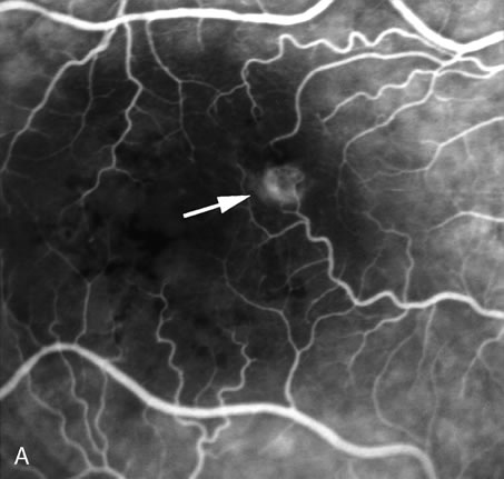
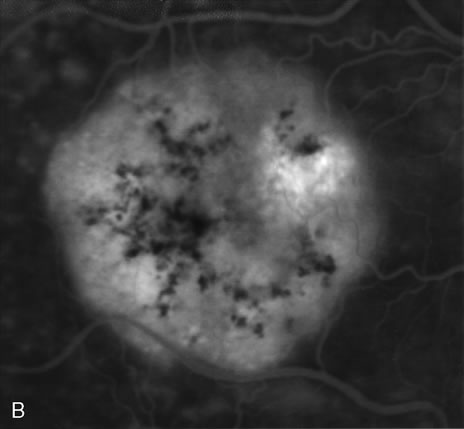
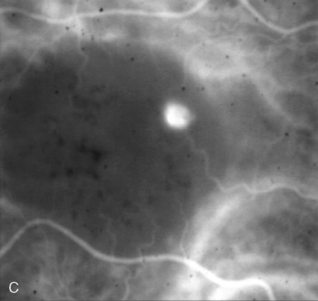
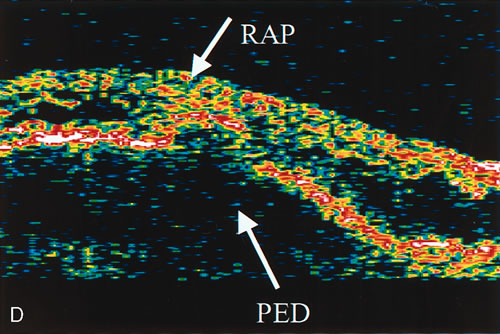
zone in senile macular degeneration. Am J Ophthalmol 93:157, 1982.

8. Yannuzzi LA, Negrão S, Iida T et al: Retinal angiomatous proliferation in age-related macular degeneration. Retina 21:416–434, 2001.

9. Fernandez LH, Freund KB, Yannuzzi LA et al: The nature of focal areas of hyperfluorescence or hot spots imaged with indocyanine green angiography. Retina 22:557–568, 2002.

10. Ghazi NG. Retinal angiomatous proliferation in age-related macular degeneration. Retina 22:509–511, 2002.

11. Axer-Siegel R, Bourla D, Priel E et al: Angiographic and flow patterns of retinal choroidal anastomoses in age-related macular degeneration with occult choroidal neovascularization. Ophthalmology 109:1726–1736, 2002.

12. Lafaut BA, Aisenbrey S, Vanden Broecke C et al: Clinicopathological correlation of deep retinal vascular anomalous complex in age related macular degeneration. Br J Ophthalmol 84:1269–1274, 2000.

13. Slakter JS, Yannuzzi LA, Schneider U: Retinal choroidal anastomoses and occult choroidal neovascularization in age-related macular degeneration. Ophthalmology 107:742–753, 2000.

14. Hartnett ME, Weiter JJ, Staurenghi G et al: Deep retinal vascular anomalous complexes in advanced age-related macular degeneration. Ophthalmology. 103:2042–2053, 1996.

15. Kuhn D, Meunier I, Soubrane G et al: Imaging of chorioretinal anastomoses in vascularized retinal pigment epithelium detachments. Arch Ophthalmol 113:1392–1398, 1995.

16. Schneider U, Gelisken F, Kreissig I: Retinal choroidal anastomosis in classic choroidal neovascularization demonstrated by indocyanine green angiography. Acta Ophthalmol Scand 73:450–452, 1995.

17. Yannuzzi LA, Ciardella A, Spaide RF et al: The expanding clinical spectrum of idiopathic polypoidal choroidal vasculopathy. Arch Ophthalmol 115:478–485, 1997.

18. Yannuzzi LA, Freund KB, Goldbaum M et al: Polypoidal choroidal vasculopathy masquerading as central serous chorioretinopathy. Ophthalmology 107:767–777, 2000.

19. Yannuzzi LA, Noguiera FB, Spaide RF et al: Idiopathic Polypoidal choroidal vasculopathy: A peripheral lesion. Arch Ophthalmol 116:382–383, 1998.

20. Yannuzzi LA, Sorenson J, Spaide RF et al: Idiopathic polypoidal choroidal vasculopathy. Retina 10:1–8, 1990.

21. Yannuzzi LA, Wong DW, Sforzolini BS et al: Polypoidal choroidal vasculopathy and neovascularized age-related macular degeneration. Arch Ophthalmol 117:1503–1510, 1999.

22. Spaide RF, Yannuzzi LA, Slakter JS et al: Indocyanine green videoangiography of idiopathic polypoidal choroidal vasculopathy. Retina 15:100–110, 1995.

23. Kleiner RC, Bruckner AJ, Johnson RL: Posterior uveal bleeding syndrome. Ophthalmology 91:110, 1984.

24. Stern RM, Zakov N, Zegarra H et al: Multiple recurrent serous sanguineous retinal pigment epithelial detachments in black women. Am J Ophthalmol 100:560–569, 1985.

25. Kleiner RC, Bruckner AJ, Johnson RL: The posterior uveal bleeding syndrome. Retina 10:9–17, 1990.

26. Perkovich BT, Zakov N, Berlin LA et al: An update on multiple recurrent serosanguineous retinal pigment epithelial detachments in black women. Retina 10:18–26, 1990.

27. MacCumber MW, Dastgheib K, Bressler NM et al: Clinicopathological correlation of the multiple recurrent serosanguineous retinal pigment epithelial detachment syndrome. Retina 15:11–110, 1995.

28. Auhja RM, Stanga PE, Vingerling JR et al: Polypoidal choroidal vasculopathy in exudative and hemorrhagic pigment epithelial detachments. Br J Ophthalmol 84:479–484, 2000.

29. Escano MF, Fujii S, Ishibashi K et al: Indocyanine green videoangiography in macular variant of idiopathic polypoidal choroidal vasculopathy. Jap J Ophthalmol 44:313–316, 2000.

30. Uyama M, Wada M, Nagai Y et al: Polypoidal choroidal vasculopathy: Natural history. Am J Ophthalmol 133:639–648, 2002.

31. Uyama M, Matsubara T, Fukushima I et al: Idiopathic polypoidal choroidal vasculopathy in Japanese patients. Arch Ophthalmol 117:1035–1042, 1999.

32. Kwok AKH, Lai TYY, Chan CWN et al: Polypoidal choroidal vasculopathy in Chinese patients. Br J Ophthalmol 86:892–897, 2002.

33. Lafaut BA, Aisenbrey S, van den Broecke C et al: Polypoidal choroidal vasculopathy pattern in age-related macular degeneration. Retina 20:650–654, 2000.

34. Lafaut BA, Leyes AM, Snyers B et al: Polypoidal choroidal vasculopathy in Caucasians. Graefes Arch Clin Exp Ophthalmol 238:752–759, 2000.

35. Lip PL, Hope-Ross MW, Gibson JM: Idiopathic polypoidal choroidal vasculopathy: A disease with diverse clinical spectrum and systemic associations. Eye 5:695–700, 2000.

36. Lois N: Idiopathic polypoidal choroidal vasculopathy in a patient with atrophic age-related macular degeneration. Br J Ophthalmol 85:1011–1012, 2001.

37. Moorthy RS, Lyon AT, Rabb MF et al: Idiopathic polypoidal choroidal vasculopathy of the macula. Ophthalmology 105:1380–1385, 1998.

38. Rosa RH Jr, Davis JL, Eifrig CW: Clinicopathologic correlation of idiopathic polypoidal choroidal vasculopathy. Arch Ophthalmol 120:502–508, 2002.

39. Ross RD, Gitter KA, Cohen C et al: Idiopathic polypoidal choroidal vasculopathy associated with retinal arterial macroaneurysm and hypertensive retinopathy. Retina 16:105–111, 1996.

40. Schneider U, Gelisken F, Inhoffen W: Clinical characteristics of idiopathic polypoidal choroid vasculopathy. Ophthalmologe 98:1186–1191, 2001.

41. Imaizumi H, Takeda M: Knobby-like choroidal neovascularization accompanied with retinal pigment epithelial detachment. Nippon Ganka Gakkai Zasshi 103:527–537, 1999.

42. Okubo A, Sameshima M, Uemara A et al: Clinicopathological correlation of polypoidal choroidal vasculopathy revealed by ultrastructural study. Br J Ophthalmol 86:1093–1098, 2002.

43. Scasellati-Sforzolini B, Mariotti C, Bryan R et al: Polypoidal choroidal vasculopathy in Italy. Retina 21:121–125, 2001.

44. Giovannini A, Amato GP, D'Altobrando E et al: Optical coherence tomography (OCT) in idiopathic polypoidal choroidal vasculopathy (IPCV). Doc Ophthalmol 97:367–371, 1999.

45. Iijima H, Iida T, Imai M et al: Optical coherence tomography of orange-red subretinal lesions in eyes with idiopathic polypoidal choroidal vasculopathy. Am J Ophthalmol 129:21–26, 2000.

46. Iijima H, Imai M, Gohodo T et al: Optical coherence tomography of idiopathic polypoidal choroidal vasculopathy. Am J Ophthalmol 127:301–305, 1999.

47. Otsuji T, Takahahashi K, Fukushima I et al: Optical coherence tomographic findings in idiopathic polypoidal choroidal vasculopathy. Ophthalmic Surg Lasers 31:210–214, 2000.

48. Quaranta M, Maget-Faysse M, Coscas G: Exudative idiopathic polypoidal choroidal vasculopathy and photodynamic therapy with verteporfin. Am J Ophthalmol 134:277–280, 2002.

49. Shiraga F, Matsuo T, Yokoe S et al: Surgical treatment of submacular hemorrhage associated with idiopathic polypoidal choroidal vasculopathy. Am J Ophthalmol 128:147–154, 1999.

50. Terasaki H, Miyake Y, Suzuki T et al: Polypoidal choroidal vasculopathy treated with macular translocation: Clinical pathological correlation. Br J Ophthalmol 86:321–327, 2002.

51. Macular Photocoagulation Study Group: Argon laser photocoagulation for neovascular maculopathy: Three-year results from randomized clinical trials. Arch Ophthalmol 104:694, 1986

52. Macular Photocoagulation Study Group: Persistent and recurrent neovascularization after krypton laser photocoagulation for neovascular lesions of age-related macular degeneration. Arch Ophthalmol 108:825, 1990.

53. Moorfields Macular Study Group: Treatment of senile disciform macular degeneration: A single-blind randomized trial by argon laser photocoagulation. Br J Ophthalmol 66:745, 1982.

54. Sorenson JA, Yannuzzi LA, Shakin JL: Recurrent subretinal neovascularization. Ophthalmology 92:1059, 1985.

55. Gass JDM: Stereoscopic Atlas of Macular Diseases: Diagnosis and Treatment. St. Louis, CV Mosby, 1987

56. Miller JW, Walsh AW, Kramer M et al: Photodynamic therapy of experimental choroidal neovascularization using lipoprotein-delivered benzoporphyrin. Arch Ophthalmol 113:8108, 1995.

57. Schmidt-Erfurth U, Miller JW, Sickenberg M et al: Photodynamic therapy with verteporfin for choroidal neovascularization caused by age-related macular degeneration: Results of retreatments in a phase 1 and 2 study. Arch Ophthalmol 117:1177–1187, 1999.

58. Miller JW, Schmidt-Erfurth U, Sickenberg M et al: Photodynamic therapy with verteporfin for choroidal neovascularization caused by age-related macular degeneration: Results of a single treatment in a phase 1 and 2 study. Arch Ophthalmol. 117:1161–1173, 1999.

59. Schmidt-Erfurth U, Miller J, Sickenberg M et al: Photodynamic therapy of subfoveal choroidal neovascularization: Clinical and angiographic examples. Graefes Arch Clin Exp Ophthalmol 236:365–374, 1998.

60. Treatment of Age-Related Macular Degeneration with Photodynamic Therapy (TAP) Study Group: Photodynamic therapy of subfoveal choroidal neovascularization in age-related macular degeneration with verteporfin: One-year results of 2 randomized clinical trials—TAP report. Arch Ophthalmol 117:1329–1345, 1999.

61. Bressler NM: Photodynamic therapy of subfoveal choroidal neovascularization in age-related macular degeneration with verteporfin: Two-year results of 2 randomized clinical trials—TAP report 2. Arch Ophthalmol. 119:198–207, 2001.

62. Bressler NM, Arnold J, Benchaboune M et al: Verteporfin therapy of subfoveal choroidal neovascularization in patients with age-related macular degeneration: Additional information regarding baseline lesion composition's impact on vision outcomes—TAP report No. 3. Arch Ophthalmol 120:1443–1454, 2002.

63. Blumenkranz MS, Bressler NM, Bressler SB et al: Verteporfin therapy for subfoveal choroidal neovascularization in age-related macular degeneration: Three-year results of an open-label extension of 2 randomized clinical trials—TAP Report No. 5. Arch Ophthalmol 120:1307–1314, 2002.

64. Lim JI: Photodynamic therapy for choroidal neovascular disease: Photosensitizers and clinical trials. Ophthalmol Clin North Am 15:473–478, 2002.

65. Spaide RF, Donsoff I, Lam DL et al: Treatment of polypoidal choroidal vasculopathy with photodynamic therapy. Retina 22:529–535, 2002.

66. Jampol LM, Scott L: Treatment of juxtafoveal and extrafoveal choroidal neovascularization in the era of photodynamic therapy with verteporfin. Am J Ophthalmol 134:99–101, 2002.

67. Spaide RF, Martin ML, Slakter J et al: Treatment of idiopathic subfoveal choroidal neovascular lesions using photodynamic therapy with verteporfin. Am J Ophthalmol 134:6–68, 2002.

68. Guidelines for using verteporfin (visudyne) in photodynamic therapy to treat choroidal neovascularization due to age-related macular degeneration and other causes. Retina . 2002 Feb;22(1):6–18. Review.

69. Dantas MA, Slakter JS, Negrao S, Fonseca RA, Kaga T, Yannuzzi LA. Photodynamic therapy with verteporfin in malatia levantinese. Ophthalmology . 2002 Feb;109(2):296–301.

70. Bressler NM. Verteporfin therapy of subfoveal choroidal neovascularization in age-related macular degeneration: two-year results of a randomized clinical trial including lesions with occult with no classic choroidal neovascularization-verteporfin in photodynamic therapy report 2. Am J Ophthalmol . 2002 Jan;133(1):168–9

71. Verteporfin in Photodynamic Therapy Report 2: Verteporfin therapy of subfoveal choroidal neovascularization in age-related macular degeneration: Two-year results of a randomized clinical trial including lesions with occult with no classic choroidal neovascularization— Am J Ophthalmol 131:541–560, 2001.

72. American Academy of Ophthalmology: Photodynamic therapy with verteporfin for age-related macular degeneration. Ophthalmology 107:2314–2317, 2000.

73. Schmidt-Erfurth U, Hasan T: Mechanisms of action of photodynamic therapy with verteporfin for the treatment of age-related macular degeneration. Surv Ophthalmol 45:195–214, 2000.

74. Gass JDM: Pathogenesis of disciform detachment of the neuroepithelium. Am J Ophthalmol 63:587, 1967.

75. Yannuzzi LA: Type-A behavior and central serous chorioretinopathy. Retina 7:111, 1987

76. Benson WE, Shields JA, Annesley WH et al: Idiopathic central serous chorioretinopathy with bullous retinal detachment. Ann Ophthalmol 12:920, 1980.

77. Yannuzzi LA, Freund KB, Goldbaum M et al: Polypoidal choroidal vasculopathy masquerading as central serous chorioretinopathy. Ophthalmology 107:767–777, 2000.

78. Gilbert CM, Owens SL, Smith PD et al: Long-term follow-up of central serous chorioretinopathy. Br J Ophthalmol 68:815, 1984.

79. Spitznas M: Central serous retinopathy. In Ryan SJ (ed): Retina, Vol 2. Medical Retina, pp 217–227. St. Louis, CV Mosby, 1989.

80. Gass JDM: Photocoagulation treatment of idiopathic central serous choroidopathy. Trans Am Acad Ophthalmol Otolaryngol 83:456, 1977.

81. Annesley WH, Augsburger JJ, Shakin JL: Ten year follow-up of photocoagulated central serous choroidopathy. Trans Am Ophthalmol Soc 79:335, 1981.

82. Roseman RL, Olk RJ: Grid laser photocoagulation for atypic central serous chorioretinopathy. Ophthalmic Surg 19:786, 1988.

83. Yannuzzi LA, Kaufman SR, Slakter J et al: Grid laser photocoagulation treatment of diffuse retinal pigment epitheliopathy (in press).

84. Klein ML, Van Buskirk M, Friedman E et al: Experience with nontreatment of central serous choroidopathy. Arch Ophthalmol 91:247, 1974.

85. Yannuzzi LA, Shakin JL, Fisher YL et al: Peripheral retinal detachments and retinal pigment epithelial atrophic tracts secondary to central serous pigment epitheliopathy. Ophthalmology 91:1554, 1984.

86. Zweng HC, Little, HL, Vassiliadis A:Argon Laser Photocoagulation, pp 117–126. St. Louis, CV Mosby, 1977.

87. Gass JDM: Bullous retinal detachment; An unusual manifestation of idiopathic central serous choroidopathy. Am J Ophthalmol 75:810–821, 1973.

88. Straatsma BR, Allen RA, Petit TH: Central serous retinopathy, Trans Pacific Coast Oto-ophthalmol Soc 47:107–127, 1966.

89. Nade, AJ, Turan, MI Coles RS: Central serous retinopathy; A generalized disease of the pigment epithelium. Mod Probl Ophthalmol 20:76–88, 1979.

90. Giovannini A: L'epitheliopathie atrophique “en trace.” Bull Mem Soc Fr Ophtalmol 94:402–406, 1982.

91. Cohen D, Gaudric A, Coscas G et al: Epitheliopathie retinienne diffuse et chorioretinopathie sereuse centrale. J Fr Ophtalmol 6:339–349, 1983.

92. von Winning CHOM, Oostheruis JA, Renger-van Dijk , AH et al: Diffuse retinal pigment epitheliopathy. Ophthalmologica, 185:7–14, 1982.

93. von Graefe, A: Ueber centrale recidivierende Retinitis. Graefes Arch Clin Exp Ophthalmol 12:211–215, 1866.

94. Bennet G: Central serous retinopathy. Br J Ophthalmol 39:605–618, 1955.

95. Maumenee AE: Symposium: Macular diseases, clinical manifestations. Trans Pacific Coast Oto-ophthalmol Soc 40:139–160, 1959.

96. Gelber GS, Schatz H: Loss of vision due to central serous chorioretinopathy following psychological stress. Am J Psychiatry 144:46–50, 1987.

97. Cordes FC: A type of foveomacular retinitis observed in the US Navy. Am J Ophthalmol 27:803, 1944.

98. Horniker, E: Su di una forma retinite centrale di origine vasoneurotica (retinite central capillaro-spastica). Ann Ottal 55:578–600, 1927.

99. Cordes, FC: A type of foveo-macular retinitis observed in the U.S. Navy. Am J Ophthalmol 27:803–816, 1944.

100. Harrington DO: The autonomic nervous system in ocular diseases. Am J Ophthalmol 29:1405–1425, 1946.

101. Harrington, DO: Psychosomatic interrelationships in ophthalmology. Am J Opthalmol 31:1241–1251, 1948.

102. Horniker E: Uber eine Form von zentraler Retinitis auf angio-neurotischer Grundlage (Retinitis centralis angio-neurotica). Graefes Arch Clin Exp Ophthalmol 123:286–360, 1929.

103. Huke J: Retinopathia centralis serosa (dissertation). Bonn, 1982.

104. Werry H, Arends C: Untersuchung zur Objektivierung von Personlichkeitsmerkmalen bei Patienten mit Retinopathia centralis serosa. Klin Monatsbl Augenheilkd 172:363–370, 1978.

105. Garg SP, Dada T, Talwar D et al: Endogenous cortisol profile in patients with central serous chorioretinopathy. Br J Ophthalmol 81:962–964, 1997.

106. Bouzas EA, Scott MH, Mastorakos G et al: Central serous chorioretinopathy in endogenous hypercortisolism. Arch Ophthalmol 111:1929–1932, 1993.

107. Jain IS, Singh K: Maculopathy, a corticosteroid side effect. J All India Ophthalmol Soc 14:250–252, 1966.

108. Eckstein MB, Spalton DJ, Holde, G: Visual loss from central serous retinopathy in systemic lupus erythematosus, Br J Ophthalmol 77:607–609, 1993.

109. Wakakura M, Ishikawa S: An evaluation of corticosteroid treatment for central serous chorioretinopathy. Rinsho Ganka 34:123–129, 1980.

110. Wakakura M, Ishikawa S: Central serous retinopathy complicating systemic corticosteroid treatment. Br J Ophthalmol 68:329–331, 1984.

111. Friberg TR, Eller, AW: Serous retinal detachment resembling central serous chorioretinopathy following organ transplantation. Graefes Arch Clin Exp Ophthalmol 288:305–309, 1990.

112. Gass JDM, Little HL: Bilateral bullous exudative retinal detachment complicating idiopathic central serous chorioretinopathy during systemic corticosteroid therapy. Ophthalmology 102:737–747, 1995.

113. Gass JDM, Slamovits TL, Fuller DG et al: Posterior chorioretinopathy and retinal detachment after organ transplantation. Arch Ophthalmol 110:1717–1722, 1992.

114. Haimoivici R, Gragoudas ES, Duker JS et al: Central serous chorioretinopathy associated with inhaled or intranasal corticosteroids. Ophthalmology 104:1653–1660, 1997.

115. Polack BCP, Baarsma GS, Snyer B: Diffuse retinal epitheliopathy complicating systemic corticosteroid treatment. Br J Ophthalmol 79:922–925, 1995.

116. Spraul CW, Lang CE, Lang GK: Central serous chorioretinopathy in systemic therapy with corticosteroids. Ophthalmologe 94:392–396, 1997.

117. Wakakura M, Song E, Ishikawa S: Corticosteroid induced central serous chorioretinopathy. Jpn J Ophthalmol 41:180–185, 1997.

118. Iida T, Spaide RF, Negrao SG et al: Central serous chorioretinopathy after epidural corticosteroid injection. Am J Ophthalmol 132:423–425, 2001.

119. Gass JD: Central serous chorioretinopathy and white subretinal exudation during pregnancy. Arch Ophthalmol 109:677–681, 1991.

120. Fastenburg, DM, Ober, RR: Central serous choroidopathy in pregnancy. Arch Ophthalmol 1055–1058, 1983.

121. Quillen, DA, Gass, JDM, Brod Rd et al: Central serous chorioretinopathy in women. Ophthalmology 103:72–79, 1996.

122. Chumbley LC, Frank RN: Central serous retinopathy and pregnancy. Am J Ophthalmol 77:15–160, 1974.

123. Bedrossian RH: Central serous retinopathy and pregnancy. Am J Ophthalmol 78:152, 1974.

124. Cruysberg JRM, Deutman AF: Visual disturbances during pregnancy caused by central serous choroidopathy. Br J Ophthalmol 66:240–241, 1982.

125. Fastenberg DM, Ober RR: Central serous choroidopathy in pregnancy. Arch Ophthalmol 101:1055–1088, 1983.

126. Sunness JS, Haller JA, Fine SL: Central serous chorioretinopathy and pregnancy. Arch Ophthalmol 111:360–364, 1993.

127. Nagayosky K: Experimental study of chorioretinopathy by intravenous injection of adrenaline, Acta Soc Ophthalmol Jpn 75:1720–1727, 1971.

128. Yoshioka H, Katsume Y, Akume H: Experimental central serous chorioretinopathy in monkey eyes: Fluorescein angiography findings. Ophthalmologica 185:168–178, 1982.

129. Yoshioka H, Sugita T, Nagayoski K: Fluorescein angiography findings in experimental retinopathy produced by intravenous adrenaline injection. Folia Ophthalmol Jpn 21:648–652, 1970

130. Yoshioka H, Katsume Y: Experimental central serous chorioretinopathy. III. Ultrastuctural findings. Jpn J Ophthalmol 26:397–409, 1982.

131. Miki T, Sunada I, Higaki T; Studies in chorioretinitis induced in stress (repeated administration of epinephrine). Acta Soc Ophthalmol Jpn 75:1037–1045, 1972.

132. Yasuzumi T, Miki T, Sugimoto K: Electron microscopic studies of epinephrine choroiditis in rabbits. I. Pigment epithelium and Bruch's membrane in the healed stage. Acta Soc Ophthalmol Jpn 78:588–598, 1974.

133. Hilton AF, Harrison JD, Lamb AM et al: Ocular complications in haemodialysis and renal transplant patients. Aust N Z J Ophthalmol 10:247–253, 1982.

134. Gass JDM: Bullous retinal detachment and multiple retinal pigment epithelium detachments in patients receiving haemodialysis. Graefes Arch Clin Exp Ophthalmol 230:454–458, 1992.

135. Cohen SM, Kokame GT, Gass JDM: Paraproteinemias associated with serous detachments of the retinal pigment epithelium and neurosensory retina. Retina 16:467–473, 1996.

136. Cunningham ET Jr, Alfred PR, Irvine AR: Central serous chorioretinopathy in patients with systemic lupus erythematosus. Ophthalmology 103:2081–2090, 1996.

137. Semon HC, Wolff E: Acute lupus erythematosus with fundus lesions. Proc Royal Soc Med 27:153–157, 1933.

138. Clifton F, Greer CH: Ocular changes in acute systemic lupus erythematosus. Br J Ophthalmol 39:1–10, 1955.

139. Burniana A: Retinal detachment in collagen disease. Ann Oculist 202:1123–1130, 1969.

140. Jabs DA, Hanneken AM, Schachat AP et al: Choroidopathy in systemic lupus erythematosus. Arch Ophthalmol 106:230–234, 1988.

141. Matsuo T, Nakayama T, Koyama T et al: Multifocal pigment epithelial damages with serous retinal detachment in systemic lupus erythematosus. Ophthalmologica 195:97–102, 1987.

142. Diddie KR, Aronson AJ, Ernest JT: Chorioretinopathy in a case of systemic lupus erythematosus. Trans Am Ophthalmol Soc 75:122–131, 1977.

143. Gold, DH, Morris, DA, Henkind, P: Ocular findings in systemic lupus erythematosus. Br J Ophthalmol 56:800–804, 1972.

144. Googe JM Jr, Brady SE, Argyle JC et al: Choroiditis in infantile periarteritis nodosa. Arch Ophthalmol 103:81–83, 1985.

145. Stefani FH, Brandt F, Pielsticker K: Periarteritis nodosa and thrombotic thrombocytopenic purpura and serous retinal detachment in siblings. Br J Ophthalmol 62:402–407, 1978.

146. Jampol LM, Lahav M, Albert DM et al: Ocular clinical findings and basement membrane changes in Goodpasture's syndrome. Am J Ophthalmol 79:452–463, 1975.

147. Kihyoun JL, Kalina RE, Klein ML: Choroidal involvement in systemic necrotizing vasculitis. Arch Ophthalmol 105:939–942, 1987.

148. Hassan L, Carvalho C, Yannuzzi LA et al: Central serous chorioretinopathy in a patient using methylenedioxymethamphetamine (MDMA) or “ecstasy.” Retina 21:559–561, 2001.

149. Klien BA: Ischemic infarcts of the choroid (Elshing's spots): A cause of retinal separation in hypertensive disease with renal insufficiency: A clinical an histopathological study. Am J Ophthalmol 66:1069–1088, 1968.

150. Stropes LL, Luft FC: Hypertensive crisis with bilateral bullous retinal detachment. JAMA 238:1948–1949, 1977.

151. Venecia G, Jampol LM: The eye in accelerated hypertension. II. Localized serous detachments of the retina in patients. Arch Ophthalmol 102:68–73, 1984.

152. Gitter KA, Houser BP, Sarin LK et al: Toxemia of pregnancy: An angiographic interpretation of fundus changes. Arch Ophthalmol 80:449–454, 1968.

153. Gass JDM, Pautler SE: Toxemia of pregnancy pigment epitheliopathy masquerading as heredomacular dystrophy. Trans Am Ophthalmol Soc 83:114–130, 1985.

154. Menchini U, Lanzetta P, Virgili G et al: Retinal pigment epithelium tear following toxemia of pregnancy. Eur J Ophthalmol 5:139–141, 1995.

155. Yamaguchi K, Abe S, Shiono T et al: Macular choroidal occlusion in dysplasmogenemia. Retina 11:423–425, 1991

156. Cogan DG: Fibrin clots in the choriocapillaris and serous detachment of the retina. Ophthalmologica 172:298–307, 1976.

157. Cogan DG: Ocular involvement in disseminated intravascular coagulopathy. Arch Ophthalmol 93:1–8, 1975.

158. Shimizu K, Tobari I: Central serous retinopathy dynamics of subretinal fluid, Mod Probl Ophthalmol 9:152–157, 1971.

159. Goldstein BG, Pavan PR: “Blow outs” in the retinal pigment epithelium. Br J Ophthalmol 71:676–681, 1987.

160. Spitznas M, Hogan MJ: Outer segments of photoreceptors and the retinal pigment epithelium: Interrelationship in the human eye. Arch Ophthalmol 84:810–819, 1970.

161. Spitznas M, Huke J: Number, shape and topography of leakage points in acute type I central serous retinopathy, Graefes Arch Clin Exp Ophthalmol 225:437–440, 1987

162. Faurschou S, Rosenberg T, Nielsen N: Central serous retinopathy and presenile disciform macular degeneration. Acta Ophthalmol 55:515–524, 1977.

163. Schatz H, Madera D, Johnson RN et al: Central serous chorioretinopathy occurring in patients 60 years of age or older. Ophthalmology, 99:63–67, 1992.

164. Berger AL, Olk RJ, Burgess, D: Central serous choroidopathy in patients over 50 years of age. Ophthalmic Surg 22:583–590, 1991.

165. Scheide, A, Nasemann JE, Lund OE: Fluorescein and indocyanine angiographies of central serous chorioretinopathy by scanning laser ophthalmoscope. Am J Ophthalmol 115:50–56, 1993.

166. Hayashi K, Hasegawa Y, Tokoro T: Indocyanine green angiography of central serous chorioretinopathy. Int Ophthalmol 9:37–41, 1986.

167. Scheider A, Hintschich C, Dimitriou S: Central serous chorioretinopathy. Studies of the site of the lesion with indocyanine green. Ophthalmologe 91:745–751, 1994.

168. Piccolino FC, Borgia L: Central serous chorioretinopathy and indocyanine green angiography. Retina 14:231–242, 1994.

169. Guyer DR, Yannuzzi, LA, Slakter JS et al: Digital indocyanine green videoangiography of central serous chorioretinopathy, Arch Ophthalmol 112:1057–1062, 1994.

170. Spaide RF, Campeas L, Haas, A et al: Central serous chorioretinopathy in younger and older adults, Ophthalmology 103:2070–2080, 1996.

171. Piccolino FC, Borgia L, Zinicola E et al: Indocyanine green angiographic findings in central serous chorioretinopathy. Eye 9:324–332, 1995.

172. Prunte C, Flammer J: Circulatory disorders of the choroid in patients with central serous chorioretinopathy. Klin Monatsbl Augenheilkd 208:337–339, 1996.

173. Lafaut BA, De Laey JJ: Indocyanine green angiography in central serous chorioretinopathy. Bull Soc Belge Ophtalmol 262:55–61, 1996.

174. Prunte C, Flammer J: Choroidal capillary and venous congestion in central serous chorioretinopathy. Am J Ophthalmol 121:26–34, 1996.

175. Iida T, Hagimura N, Otani T et al: Choroidal vascular lesions in serous retinal detachment viewed with indocyanine green angiography. Nippon Ganka Gakkai Zasshi 100:817–824, 1996.

176. Menchini U, Virgili G, Lanzetta P et al: Indocyanine green angiography in central serous chorioretinopathy. Int Ophthalmol 21:57–69, 1997.

177. Giovannini A, Scasellati Sforzolini B, D'Altobrando E: Choroidal findings in the course of idiopathic serous pigment epithelium detachment detected by indocyanine green videoangiography. Retina 17:286–293, 1997.

178. Okushiba U, Takeda M: Study of choroidal vascular lesions in central serous chorioretinopathy using indocyanine green angiography. Nippon Ganka Gakkai Zasshi 101:74–82, 1997.

179. Spaide, RF, Orlock D, Herrmann-Delemazure B et al: Wide-angle indocyanine green angiography. Retina 18:44–49, 1998.

180. Guyer DR: Central serous chorioretinopathy. In Yannuzzi LA, Flower RW, Slakter JS (eds): Indocyanine Green Angiography, pp 297–304. St. Louis, Mosby –Year Book, 1997.

181. Spaide RF, Hall L, Haas A: Indocyanine green videoangiography of central serous chorioretinopathy in older adults. Retina 16:78–80, 1996.

182. Nanjian, M: Long-term follow-up of central serous retinopathy, Trans Ophthalmol Soc UK 97:656–661, 1977.

183. Dellaporta A: Central serous retinopathy. Trans Am Ophthalmol Soc 74:144–153, 1976.

184. Watzke RC, Burton TC, Woolson RF: Direct and indirect laser photocoagulation of central serous choroidopathy, Am J Ophthalmol 88:914–918, 1979.

185. Wessing A: Zur Pathogenese und Therapie der sogenannten Retinitis centralis serosa. Ophthalmologica 153:259–276, 1967.

186. Wessing A: Die Therapie der Retinitis centralis serosa mit Lichtcoagulation. Berl Dtsch Ophthalmol Ges 68:429–431, 1968.

187. Wessing A: Changing concepts of central serous retinopathy and its treatment. Trans Am Acad Ophthalmol Otolaryngol 77:275–280, 1973.

188. Watzke RC, Burton TC, Leaverton PA: Ruby laser photocoagulation therapy of central serous retinopathy. Trans Am Acad Ophthalmol Otolaryngol 78:205–211, 1974.

189. L'Esperance FA Jr: Argon and ruby laser photocoagulation of disciform macular disease. Trans Am Acad Ophthalmol Otolaryngol 75:609–625, 1971.

190. Spalter HF: Photocoagulation of central serous retinopathy: A preliminary report. Arch Ophthalmol 79:247–253, 1968.

191. Annesley WH Jr, Augsburger JJ, Shakin JL: Ten-year follow-up of photocoagulated central serous choroidopathy. Trans Am Ophthalmol Soc 79:335–346, 1981.

192. Leaver P, Williams C: Argon laser photocoagulation in the treatment of central serous retinopathy. Br J Ophthalmol 63:674–677, 1979.

193. Ficker L, Vafadis G, While A et al: Long term results of treatment of central serous retinopathy. Trans Ophthalmol Soc UK 105:473–475, 1986.

194. Ficker L, Vafadis G, While A et al: Long term follow-up of a prospective trial of argon laser photocoagulation in the treatment of central serous retinopathy. Br J Ophthalmol 72:829–834, 1988.

195. Yannuzzi LA, Slakter JS, Kaufman SR et al: Laser treatment of diffuse retinal pigment epitheliopathy. Eur J Ophthalmol, 2:103–114, 1992.

196. Piccolino FC: Laser treatment of eccentric leaks in central serous chorioretinopathy resulting in disappearance of untreated juxtafoveal leaks. Retina 12:96–102, 1992.

197. L'Esperance, FA: Ocular Photocoagulation, pp 190–197. St. Louis, Mosby–Year Book, 1975.

198. Robertson DM, Ilstrup D: Direct, indirect and sham laser photocoagulation in the management of sham central serous chorioretinopathy. Am J Ophthalmol 94:457–466, 1983.

199. Eng-Yiat Y, Robertson DM: The long term outcome of central serous chorioretinopathy. Arch Ophthalmol 114:689–692, 1996.

200. Brancato R, Scialdone A, Pece A et al: Eight-year follow-up of central serous chorioretinopathy with and without laser treatment. Graefes Arch Clin Exp Ophthalmol 225:166–168, 1987.

201. Annesley VH Jr, Augsburger JJ, Shakin JL: Ten-year follow-up of photocoagulated central serous choroidopathy. Trans Am Ophthalmol 79:335–346, 1981.

202. Castro-Correia J, Coutinho MF, Rosas V: Long term follow up of central serous chorioretinopathy in 150 patients. Doc Ophthalmol 81:379–386, 1992.

203. Schatz H, Yannuzzi LA, Gitter KA: Subretinal neovascularization following argon laser photocoagulation treatment for central serous chorioretinopathy; complication or misdiagnosis? Trans Am Acad Ophthalmol Otolaryngol 83:893–906, 1977.

204. Schatz, H, McDonald HR, Johnson RN et al: Subretinal fibrosis in central serous chorioretinopathy, Ophthalmology, 102:1077–1088, 1995.

205. Schatz H, D'Oesterloh M, McDonald RH et al: Development of retinal vascular leakage and cystoid macular edema secondary to central serous chorioretinopathy. Br J Ophthalmol 77:744–746, 1993.

206. Weiler W, Foerester MH, Wessing A: Exudative retinal detachment, pigment epithelium tear and subretinal exudate in a case of central serous chorioretinopathy. Klin Monatsbl Augenheilkd 199:450–453, 1991.

207. McDonald HR, Schatz H: Introduction to epiretinal membranes. In Ryan SJ (ed): Retina, Vol 2. Medical Retina. St. Louis, CV Mosby, 1989.

208. Margherio RR, Cox MS, Trese MT et al: Removal of epimacular membranes. Ophthalmology 92:1075, 1985.

209. Rice TA, de Bustros S, Michels RG et al: Prognostic factors in vitrectomy for epiretinal membranes of the macula. Ophthalmology 93:602, 1986.

210. Green RL, Byrne SF: Diagnostic ophthalmic ultrasound. In Ryan SJ (ed): Retina, Vol 1. Basic Science and Inherited Retina Disease, pp 191–273. St. Louis, CV Mosby, 1989.

211. Sidd RJ, Fine SL, Owens SL et al: Idiopathic preretinal gliosis. Am J Ophthalmol 94:44, 1982.

212. Foos RY: Vitreoretinal juncture: Epiretinal membranes and vitreous. Invest Ophthalmol Vis Sci 16:416, 1977.

213. McDonnell PJ, Fine SL, Hillis Al: Clinical features of idiopathic macular cysts and holes. Am J Ophthalmol 93:777, 1982,

214. Morgan CM, Schatz H: Idiopathic macular holes. Am J Ophthalmol 99:437, 1985,

215. Hee MR, Puliafito CA, Wong C et al: Optical coherence tomography of macular holes. Ophthalmology 102:748–756, 1995.

216. Dai H, Li Y, Long L et al: The observation on detachment of posterior hyaloid membrane in idiopathic macular holes by optical coherence tomography. Chung Hua Yen Ko Tsa Chih 38:667–669, 2002.

217. Apostolopoulos MN, Koutsandrea CN, Moschos MN et al: Evaluation of successful macular hole surgery by optical coherence tomography and multifocal electroretinography. Am J Ophthalmol 134:667–674, 2002.

218. Uemoto R, Yamamoto S, Aoki T et al: Macular configuration determined by optical coherence tomography after idiopathic macular hole surgery with or without internal limiting membrane peeling. Br J Ophthalmo. 86:1240–1242, 2002.

219. Ullrich S, Haritoglou C, Gass C et al: Macular hole size as a prognostic factor in macular hole surgery. Br J Ophthalmol 86:390–393, 2002.

220. Ip MS, Baker BJ, Duker JS et al: Anatomical outcomes of surgery for idiopathic macular hole as determined by optical coherence tomography. Arch Ophthalmol 120:29–35, 2002.

221. Tanner V, Chauhan DS, Jackson TL et al: Optical coherence tomography of the vitreoretinal interface in macular hole formation. Br J Ophthalmol 85:1092–1097, 2001.

222. Haouchine B, Massin P, Gaudric A: Foveal pseudocyst as the first step in macular hole formation: A prospective study by optical coherence tomography. Ophthalmology 108:15–22, 2001.

223. Jumper JM, Gallemore RP, McCuen BW 2nd et al: Features of macular hole closure in the early postoperative period using optical coherence tomography. Retina 20:232–237, 2000.

224. Spiritus A, Dralands L, Stalmans P et al: OCT study of fellow eyes of macular holes. Bull Soc Belge Ophtalmol 275:81–84, 2000.

225. Gallemore RP, Jumper JM, McCuen BW 2nd et al: Diagnosis of vitreoretinal adhesions in macular disease with optical coherence tomography. Retina 20:115–120, 2000.

226. Akasaka Y, Nishikawa S, Tamai M: Analysis of the retinal edema of full-thickness macular holes by scanning laser ophthalmoscopy and optical coherence tomography. Tohoku J Exp Med 189:233–238, 1999.

227. Ripandelli G, Coppe AM, Bonini S et al: Morphological evaluation of full-thickness idiopathic macular holes by optical coherence tomography. Eur J Ophthalmol 9:212–216, 1999.

228. Stalmans P, Spileers W, Dralands L: The use of optical coherence tomography in macular diseases. Bull Soc Belge Ophtalmol 272:15–30, 1999.

229. Mori K, Abe T, Yoneya S: Vitreoretinal tomography and foveolar traction in macular hole development and macular pseudohole. Nippon Ganka Gakkai Zasshi 103:371–378, 1999.

230. Pal E, Givort G, Laroche A et al: Macular imaging with optical coherence tomography. J Fr Ophtalmol 21:484–494, 1998.

231. Smiddy WE, Michels RG, Glaser BM et al: Vitrectomy for impending idiopathic macular holes. Am J Ophthalmol 105:371, 1988.

232. Jost BF, Hutton WL, Fuller DG et al: Vitrectomy in eyes at risk for macular hole formation. Ophthalmology 97:843, 1990.

233. Akiba J, Yoshida A, Trempe CL: Risk of developing a macular hole. Arch Ophthalmol 108:1088, 1990.

234. Margherio RR, Schepens CL: Macular breaks: I. Diagnosis, etiology, and observations. Am J Ophthalmol 74:219, 1972.

235. Morgan CM, Schatz H: Involutional macular thinning. Ophthalmology 93:153, 1986.

236. Guyer DR, Green WR, de Bustros S et al: Histopathologic features of idiopathic macular holes and cysts. Ophthalmology 97:1045, 1990.

237. Avila MP, Jalkh AE, Murakami K et al: Biomicroscopic study of the vitreous in macular breaks. Ophthalmology 90:1277, 1983.

238. Johnson RN, Gass JDM: Idiopathic macular holes: Observations, stages of formation, and implications of surgical intervention. Ophthalmology 95:917, 1988.

239. Gass JDM, Anderson DR, Davis EB: A clinical, fluorescein angiographic, and electron microscopic correlation of cystoid macular edema. Am J Ophthalmol 100:82, 1985.

240. Flach AJ, Dolan BJ, Irvine AR: Effectiveness of ketorolac tromethamine 0.5% ophthalmic solution for chronic aphakic and pseudophakic cystoid macular edema. Am J Ophthalmol 103:479, 1987.

241. Cox SN, Hay E, Bird AC: Treatment of chronic macular edema with acetazolamide. Arch Ophthalmol 106:1190, 1988.

242. Fishman GA, Gilbert LD, Fiscella RG et al: Acetazolamide for treatment of chronic macular edema in retinitis pigmentosa. Arch Ophthalmol 107:1445, 1989.

243. Zierhut M, Thiel HJ, Schlote T: Treatment of uveitic macular edema with acetazolamide. Doc Ophthalmol 97:409–413, 1999.

244. Wolfensberger TJ: The role of carbonic anhydrase inhibitors in the management of macular edema. Doc Ophthalmol 97:387–397, 1999.

245. Flach AJ. The incidence, pathogenesis and treatment of cystoid macular edema following cataract surgery. Trans Am Ophthalmol Soc 96:557–634, 1998.

246. Moldow B, Sander B, Larsen M et al: The effect of acetazolamide on passive and active transport of fluorescein across the blood-retina barrier in retinitis pigmentosa complicated by macular oedema. Graefes Arch Clin Exp Ophthalmol 236:881–889, 1998.

247. Grover S, Fishman GA, Fiscella RG et al: Efficacy of dorzolamide hydrochloride in the management of chronic cystoid macular edema in patients with retinitis pigmentosa. Retina 17:222–231, 1997.

248. Rho D: Acetazolamide treatment of CME in patients with uveitis. Ophthalmology 103:1717, 1996. Flach A. Acetazolamide treatment of CME in patients with uveitis. Ophthalmology 103:1715–1716, 1996.

249. Whitcup SM, Csaky KG, Podgor MJ et al: A randomized, masked, cross-over trial of acetazolamide for cystoid macular edema in patients with uveitis. Ophthalmology 103:1054–1062, 1996.

250. Horgan SE, Fraser SG, Ferrante PF et al: Cystoid macular oedema in chronic myeloid leukaemia: treatment with acetazolamide and response to bone marrow transplantation. Eye 10:394–6, 1996.

251. Avila MP, Weiter JJ, Jalkh AE et al: Natural history of choroidal neovascularization in degenerative myopia. Ophthalmology 91:1573, 1984.

252. Brancato R, Pece A, Avanza P, Radrizzani E: Photo-coagulation scar expansion after laser therapy for choroidal neovascularization in degenerative myopia. Retina 10:239, 1990.

253. Soubrane G, Pison J, Bornert P et al: Néovaisseaux sous-rétiniens de la myopic dégénérative: Résultats de la photo-coagulation. Bull Soc Ophtalmol Fr 86:269, 1986.

254. Rabb MF, Garoon I, LaFranco FP: Myopic macular degeneration. Int Ophthalmol Clin 21:51, 1981.

255. Avetisov ES, Savitskaya NF: Some features of ocular microcirculation in myopia. Ann Ophthalmol 9:1261, 1977.

256. VIP Report No. 1: Photodynamic therapy of subfoveal choroidal neovascularization in pathologic myopia with verteporfin. 1-year results of a randomized clinical trial.— Ophthalmology 108:841–852, 2001.

257. Dean Hart JCD, Frank HJ: Retinal opacification after blunt non-perforating concussional injuries to the globe. Trans Ophthalmol Soc UK 95:94, 1975.

258. Burton TC: Unilateral Purtscher's retinopathy. Ophthalmology 87:1096, 1980.

259. Grant WM: Toxicology of the Eye, 3rd ed. Springfield, IL, Charles C Thomas, 1986.

260. Cruess AF, Schachat AP, Nicholl J et al: Chloroquine retinopathy: Is fluorescein angiography necessary? Ophthalmology 92:1127, 1985.

261. Easterbrook M: The use of Amsler grids in early chloroquine retinopathy. Ophthalmology 91:1368, 1984.

262. Bullock JD, Albert DM: Flecked retina: Appearance secondary to oxalate crystals from methoxyflurane anesthesia. Arch Ophthalmol 93:26, 1975.

263. Meredith TA, Wright JD, Gammon JA et al: Ocular involvement in primary oxalosis. Arch Ophthalmol 102:584, 1984.

264. Fielder AR, Garner A, Chambers TL: Ophthalmic manifestations of primary oxalosis. Br J Ophthalmol 64:782, 1980.

265. Mackool RJ, Muldoon T, Fortier A et al: Epinephrine-induced cystoid macular edema in aphakic eyes. Arch Ophthalmol 95:791, 1977.

266. McKeown CA, Swartz M, Blom J et al;: Tamoxifen retinopathy. Br J Ophthalmol 65:177, 1981.

267. Craythorn JM, Swartz M, Creel DJ: Clofazimine-induced bull's-eye retinopathy. Retina 6:50, 1986.

268. Cunningham CA, Friedberg DN, Carr RE: Clofazimine-induced generalized retinal degeneration. Retina 10:131, 1990.

269. Balian JV: Accidental intraocular tobramycin injection: A case report. Ophthalmic Surg 14:353, 1983.

270. McDonald HR, Schatz H, Allen AW et al: Retinal toxicity secondary to intraocular gentamicin injection. Ophthalmology 93:871, 1986.

271. Swartz M: Other diseases: Drug toxicity and metabolic and nutritional conditions. In Ryan SJ (ed): Retina, Vol 2. Medical Retina, pp 737–748. St. Louis, CV Mosby, 1989.

272. Tso MOM, La Piana FG: The human fovea after sungazing. Trans Am Acad Ophthalmol Otolaryngol 79:788, 1975.

273. Penner R, McNair JN: Eclipse blindness: Report of an epidemic in the military population of Hawaii. Am J Ophthalmol 61:1452, 1966.

274. Freedman J, Gombos GM: Fluorescein fundus angiography in self-induced solar retinopathy: A case report. Can J Ophthalmol 6:124, 1971.

275. McDonald HR, Irvine AR: Light-induced maculopathy from the operating microscope in extracapsular cataract extraction and intraocular lens implantation. Ophthalmology 90:945, 1983.

276. Khwarg SG, Geoghegan M, Hanscom TA: Light-induced maculopathy from the operating microscope. Am J Ophthalmol 98:628, 1984.

277. Calkins JL, Hochheimer BF: Retinal light exposure from operating microscopes. Arch Ophthalmol 97:2363, 1979.

278. Mannis MJ, Becker B: Retinal light exposure and cystoid macular edema. Arch Ophthalmol 98:1133, 1980.

Back to Top