|
|
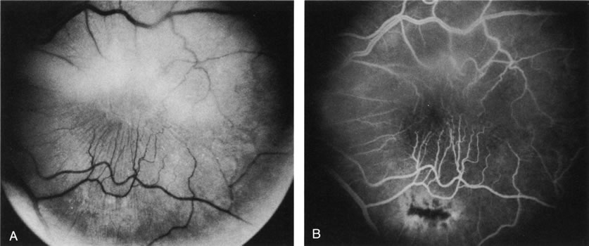
| Fig. 32. Preretinal gliosis. A. Red-free photograph demonstrates stretching of retinal vessels in the inferior macula toward the inferior arcade. The preretinal membrane apparently derives from a dense condensation of fibrous tissue in the inferior macula. The superior half of the macula is partly obscured by a fibrous condensation from posterior vitreous detachment. B. Fluorescein angiogram also demonstrates the traction on the retinal vessels, and the dense gliotic membrane partially blocks background fluorescence. There is some mild telangiectasia of the perifoveal retinal capillaries. |