|
|
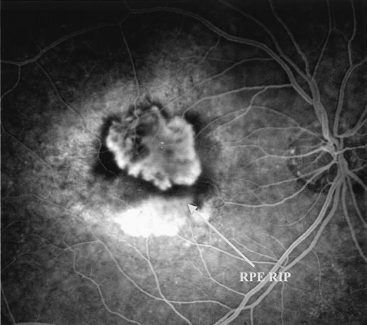
| Fig. 6. Classic, submacular choroidal neovascularization (CNV) and retinal pigment epithelium (RPE) rip. Fluorescein angiography reveals the presence of a well-defined subfoveal CNV and hyperfluorescent area in the inferior macula corresponding to the RPE rip (yellow arrow). |