|
|
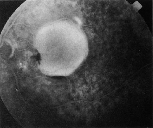
| Fig. 4. Subretinal blood from a choroidal neovascular membrane. A small hemorrhage has layered out at the inferior aspect of a large retinal pigment epithelium (RPE) detachment. A shallow overlying neurosensory detachment can be appreciated as the slightly darkened, narrow band that surrounds the RPE detachment. The neurosensory detachment is being filled with fluorescein through a break in the RPE. The subretinal neovascular membrane, not clearly evident in this early-phase fluorescein angiogram, is at the nasal edge of the RPE detachment, near the optic disc. |