|
|
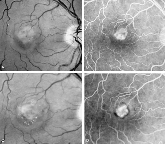
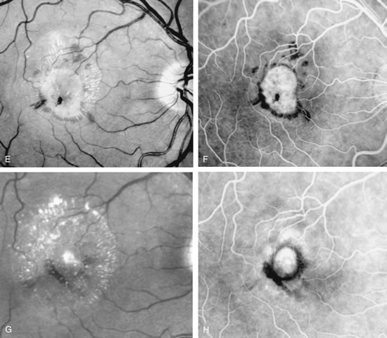
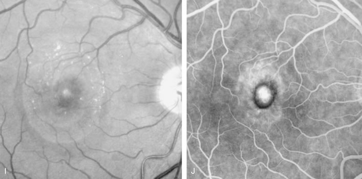
| Fig. 20. A. Red-free photograph of a 20-year-old patient with sudden loss of vision to the level of 20/200. There is exudative, neurosensory macular detachment, a few hemorrhages, and lipid exudates. B. Fluorescein angiography reveals the presence of classic choroidal neovascularization (CNV), which appears to be juxtafoveal (<200 μ from fixation). Given the size of the CNV and its proximity to the fovea, it was decided to treat the patient with photodynamic treatment (PDT). C. Red-free photograph of the same eye 2 weeks after PDT; there is increased subretinal exudation D. Fluorescein angiography demonstrates that the CNV is still actively leaking. E. Red-free photograph 4 weeks after PDT demonstrates further increase in the size of the neurosensory macular detachment, subretinal hemorrhages, and lipid exudation. F. Fluorescin angiography reveals that the CNV has extended under the fovea. Given the young age of the patient, an inflammatory component of the neovascular process was suspected. It was decided to give a posterior, subtenon injection of triamcinolone acetonide, 40 mg/1 mL. G. Two weeks after steroid treatment there is partial reabsorption of the subretinal fluid. H. Fluorescein angiography demonstrates contraction of the CNV. I. Four weeks after injection of triamcinolone there is further reduction in the degree of neurosensory detachment; vision had improved to 20/60. J. Fluorescein angiography demonstrates that the CNV is smaller and less active (less leakage). |