|
|
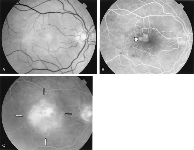
| Fig. 11. A. Red-free photograph of the right eye of a patient with wet age-related macular degeneration reveals exudative, neurosensory detachment in the macula and a few subretinal hemorrhages. B. Early-phase fluorescein angiography demonstrates well-defined classic choroidal neovascularization (CNV) (arrowhead). C. Late-phase fluorescein angiography shows leakage of dye from the classic CNV surrounded by an area of late hyperfluorescence consistent with occult CNV (arrows). |