|
|
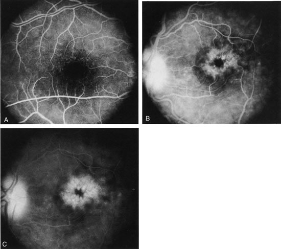
| Fig. 36. Cystoid macular edema (CME). This patient had bilateral vitritis and CME. A. Early-phase photograph of the right eye reveals telangiectasia of the perifoveal retinal capillaries with some early leakage visible temporally. B. Mid-phase photograph of the left eye reveals more intense fluorescence leakage. C. Late-phase photograph of the left eye demonstrates cystic accumulation of fluorescein in a classic “petaloid” configuration. The late-phase staining of the optic nerve head in this fluorescein angiogram suggests an inflammatory cause of the CME. |