|
|
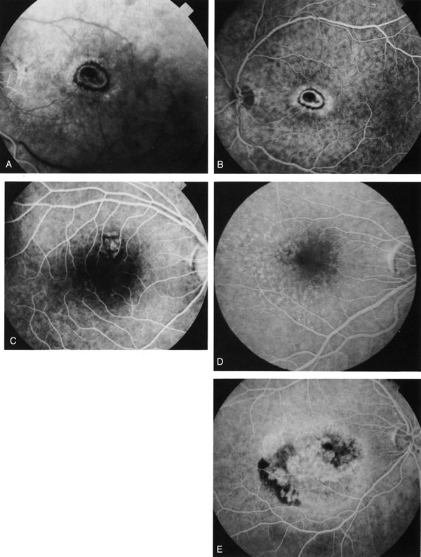
| Fig. 16. Subfoveal, juxtafoveal, and extrafoveal choroidal neovascular membranes. A and B. Large subfoveal choroidal neovascularization (CNV) in a 69-year-old man with blood and pigment blocking central fluorescence on both the early-phase (A) and late-phase (B) photographs. The hypofluorescence surrounding the membrane is commonly seen in CNV and may be due to lipofuscin. C. Juxtafoveal CNV in a 37-year-old man with idiopathic CNVM. D. Cuticular drusen in same patient as in C were asymptomatic. E. Years later, this same patient developed a large extrafoveal CNV with central macular pigment abnormalities. A large neurosensory detachment was responsible for the disappearance of the drusen. (Courtesy of Dr. Kenneth G. Noble.) |