|
|
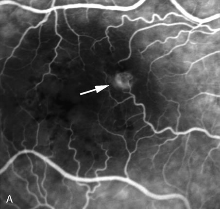
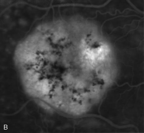
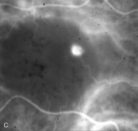
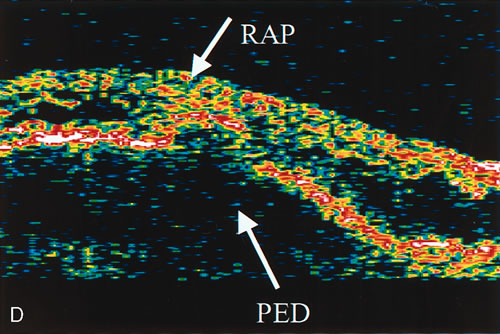
| Fig. 13. A. Early-phase Fluorescein angiography demonstrating the presence of an intraretinal angiomatous lesion (arrow). There is an associated pigment epithelium defect (PED), which is still hypofluorescent. B. Late-phase fluorescein angiography shows leakage from the retinal angiomatous proliferation (RAP) lesion and polling of dye into the PED. C. Indocyanine green angiogram of the same eye better demonstrates the presence of a hot spot corresponding to the RAP lesion. The PED remains hypofluorescent. D.Optical clearance tomography image demonstrates the presence of a serous PED and of intraretinal neovascularization. |