|
|
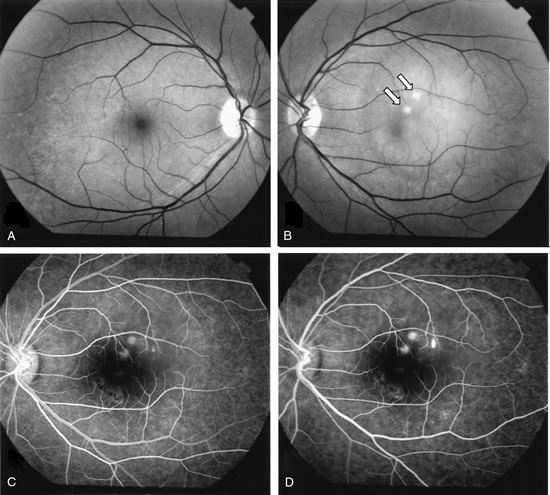
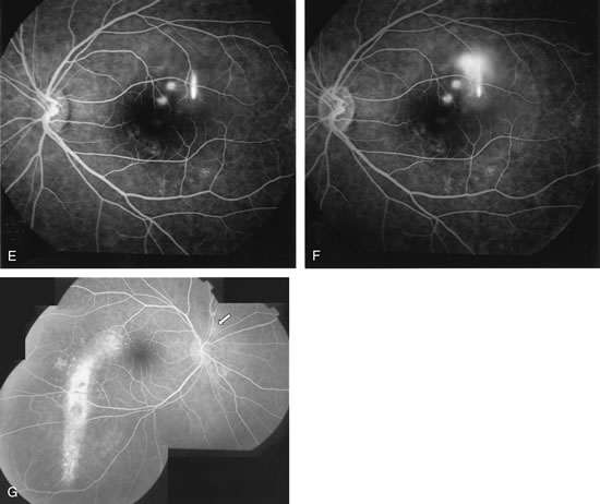
| Fig. 25. A 41-year-old man with the diagnosis of central serous chorioretinopathy in both eyes for 10 years presented complaining of sudden decrease of vision in the left eye. A. Red-free photograph of the right eye reveals pigmentary changes temporal to the fovea. B. Red-free photograph of the left eye shows a well-circumscribed neurosensory detachment of the macula with two areas of focal pigment epithelium detachment (PED). C– F. Fluorescein angiography shows two localized areas of PED and the typical “smokestack” appearance of the dye leaking under the detached retina. Note that the dye expands in an umbrella-like fashion once it reaches the upper limit of the detachment. G. Mid-phase fluorescein angiography of the fellow eye shows window-defect hyperfluorescence corresponding to the retinal pigment epithelium tract that extends inferiorly. |