|
|
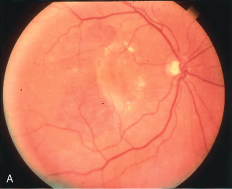
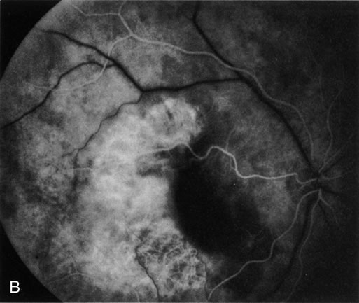
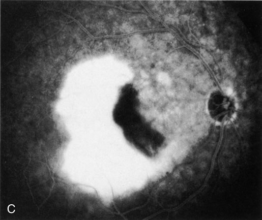
| Fig. 5. A. Clinical photograph of a large, crescent-shaped rip of the retinal pigment epithelium (RPE) in the temporal macula. B. Early-phase fluorescein angiography study demonstrates the presence of a window defect corresponding to the RPE rip, which exposes the choroidal vasculature. Where the RPE is redundant in the central macula there is blockage of the normal choriocapillaris fluorescence. C. Late-phase angiogram reveals intense hyperfluorescence seen through the RPE defect. |