|
|
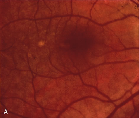
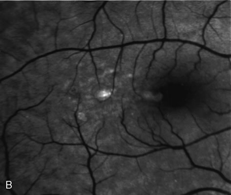
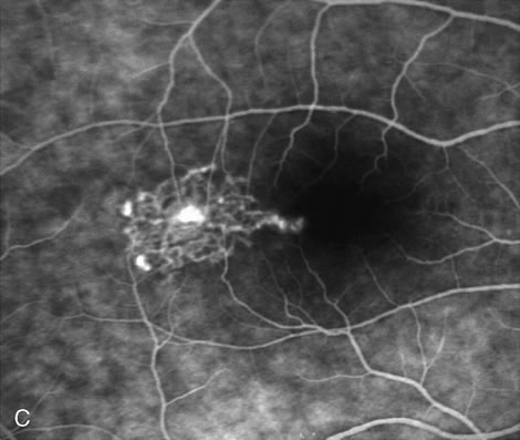
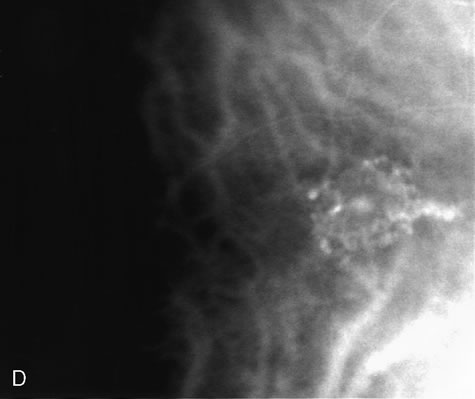
| Fig. 15. A 51-year-old Caucasian woman was referred with diagnosis of central serous chorioretinopathy in her right eye. A and B. Color photograph and red-free photograph of the right eye show the presence of atrophic changes in the retinal pigment epithelium consistent with central serous chorioretinopathy. C, Fluorescein angiogram of the right eye reveals the presence of avascular network of small-caliber vessels terminating in polyp-like structures. D. Indocyanine green angiogram of the right eye confirms the presence of the polypoidal lesion. |