|
|
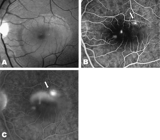
| Fig. 24. A 34-year-old Caucasian man presented, complaining of blurred vision in his left eye of 1-week duration. A. Clinical photograph of the left eye shows serous neurosensory macular detachment. B and C. Fluorescein angiography demonstrates a pinpoint area of hyperfluorescence in the central macula, leading to the characteristic smokestack configuration seen in the late-phase angiogram. A pigment epithelial detachment temporal to the fovea is increasing in hyperfluorescence throughout the study (white arrows). |