|
|
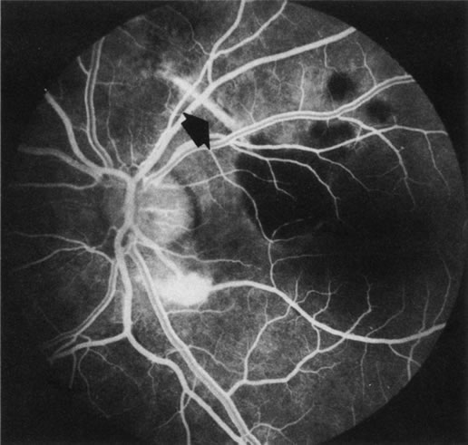
| Fig. 39. Choroidal rupture. A large choroidal rupture arcs circumferentially over the optic nerve head (arrow). The hyperfluorescence is from scleral staining that is easily visible through this defect of the retinal pigment epithelium, Bruch's membrane, and choriocapillaris. There is blocked fluorescence where laser treatment has obliterated a choroidal neovascularization that developed at the inferior aspect of the defect. (Courtesy of Dr. Kenneth G. Noble.) |