|
|
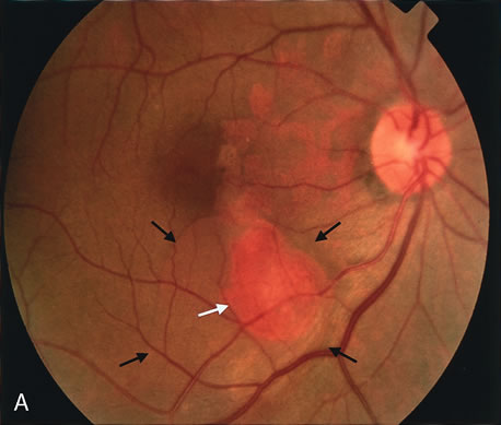
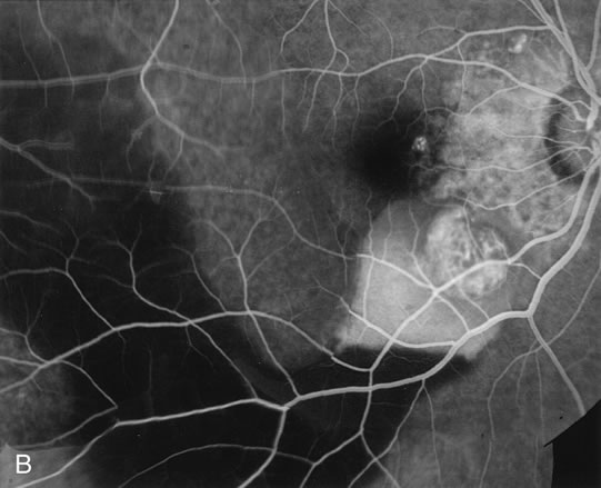
| Fig. 14. A. Color photograph of the right eye shows a ramified pattern of choroidal vascular abnormality irradiating from the peripapillary area toward the macula. The dilated vascular channels end with bulging polyp-like structures. A larger, orange, saccular dilation is seen inferior to the macula (white arrow); leakage of fluid from this vascular abnormality results in serosanguineous pigment epithelium detachment (black arrows). B. The corresponding fluorescein angiogram composite highlights the vascular lesion in the peripapillary area and the serosanguineous detachment of the pigment epithelium that extends inferiorly and temporally off the macula. |