
|
|
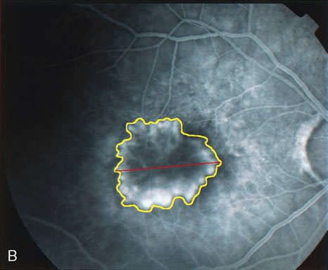
| Fig. 19. A. Red-free photograph of the right eye of a patient with exudative angiomatous macular degeneration. B. Fluorescein angiography reveals the presence of subfoveal, classic choroidal neovascularization (CNV). The boundaries of the CNV are digitally traced (yellow), and the greater linear dimension of the lesion is measured (red) to guide the PDT. |