|
|
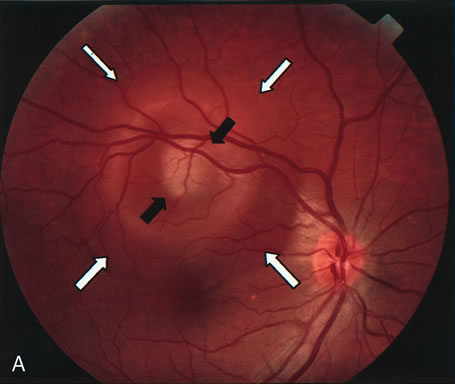
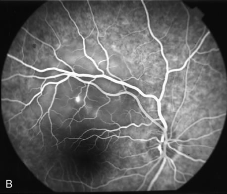
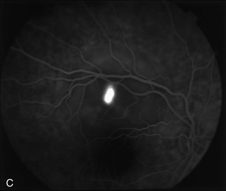
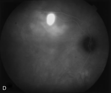
| Fig. 26. A 36-year-old man presented with decreased visual acuity in the right eye. A. Color photograph of the right eye shows serous neurosensory detachment (white arrows) in the superior area of the macula with ring-like yellowish subretinal nodular deposits consistent with fibrin surrounding the localized pigment epithelium detachment (PED) (black arrows). B. Early-phase fluorescein angiography reveals a localized area of leakage corresponding to serous PED. C. Late-phase fluorescein angiography demonstrates hyperfluorescence due to pooling beneath the serous PED. D. Late-phase indocyanine green angiogram shows an area of hyperfluorescence corresponding to the serous PED with staining of the subretinal fibrin deposits. |