|
|
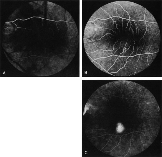
| Fig. 22. Central serous chorioretinopathy. A. In this arterial-phase photograph, a small area of early hyperfluorescence represents a small RPE detachment in the inferotemporal macula. B. This mid-phase photograph reveals increased fluorescence in the area, reflecting filling of a neurosensory detachment. C. The detachment continues to fill with fluorescence in this late-phase photograph. This gradually enlarging fluorescence is sometimes called “ink blot” filling of the subretinal space. (Courtesy of Dr. Kenneth G. Noble.) |