|
|
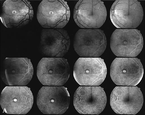
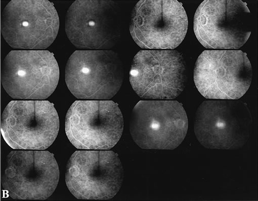
| Fig. 7. Composite photograph of fluorescein angiography study in a patient with classic, subfoveal choroidal neovascularization (CNV) in the right eye. A The classic neovascular membrane appears as a well-defined area of hyperfluorescence in the early phases of the angiogram. There is leakage of dye from the classic net in the subretinal space throughout the study. B. In the late phase of the study, the edges of the CNV are fuzzy and indistinguishable. |