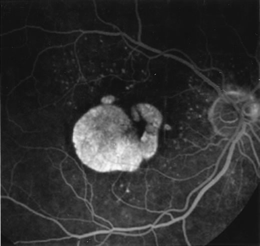
Fig. 1.
Late-phase fluorescein angiography of an eye with a central area of geographic atrophy, which appears hyperfluorescent. A few soft drusen are also present.