|
|
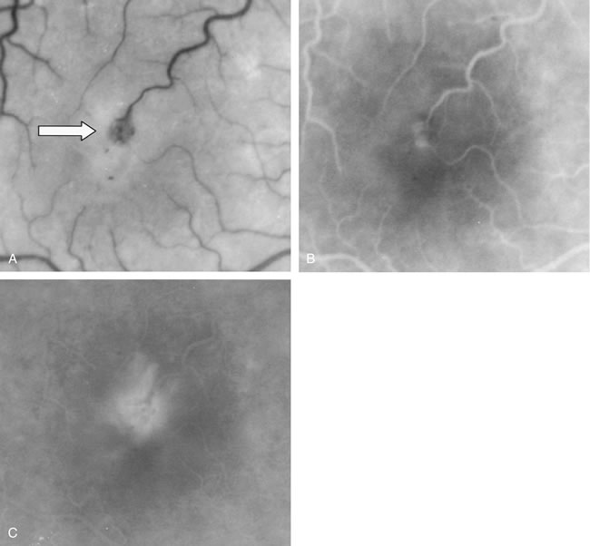
| Fig. 12. A. Clinical photograph of a retinal angiomatous proliferation (RAP) lesion (arrow). Note the intraretinal angiomatous proliferation, a feeding retinal arteriole, and a draining retinal venule, as well as the presence of intraretinal hemorrhages. B–C. Fluorescein angiography reveals late leakage from the RAP lesion. |