|
|
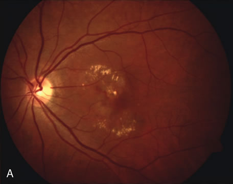
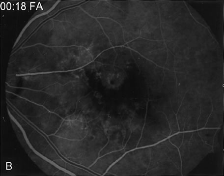
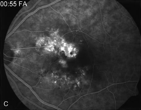
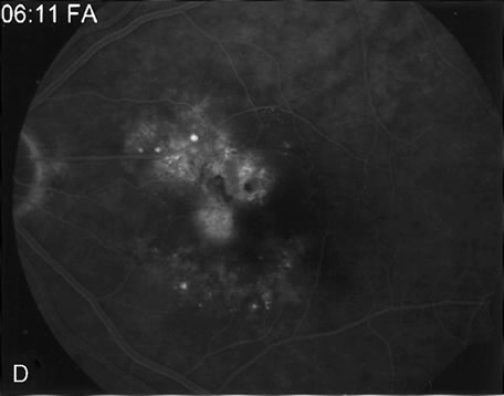
| Fig. 9. A. Clinical photograph of the left eye of a patient with exudative neurosensory macular detachment. There were also intraretinal and subretinal hard exudates, subretinal hemorrhage, and retinal pigment epithelium (RPE) changes. B–D. Fluorescein angiography of the same eye demonstrates the presence of stippled hyperfluorescence from the RPE, and late-phase oozing of dye of undefined origin. There was occult choroidal neovascularization. |