Chapter 110a
Diagnostic Indocyanine Green Angiography: Clinical Applications
NICOLE E. GROSS, JASON S. SLAKTER, LINDSAY M. SMITHEN, LAWRENCE A. YANNUZZI, CHRISTINA M. KLAIS, JOHN A. SORENSON, RICHARD F. SPAIDE, YALE L. FISHER and DENNIS A. ORLOCK
Main Menu   Table Of Contents

Search

OCCULT CHOROIDAL NEOVASCULARIZATION
CHOROIDAL INFLAMMATORY DISEASE
BIRDSHOT CHORIORETINOPATHY
MULTIPLE EVANESCENT WHITE DOT SYNDROME
ACUTE MULTIFOCAL POSTERIOR PLACOID PIGMENT EPITHELIOPATHY
SERPIGINOUS CHORIORETINOPATHY
HARADA DISEASE
MULTIFOCAL CHOROIDITIS
OCULAR HISTOPLASMOSIS SYNDROME
CENTRAL SEROUS CHORIORETINOPATHY
IDIOPATHIC POLYPOIDAL CHOROIDAL VASCULOPATHY
CHOROIDAL TUMORS
CONCLUSIONS
REFERENCES

The special properties of indocyanine green (ICG) dye and the advent of digital imaging techniques have provided for enhanced imaging of the choroid for more accurate and specific diagnoses and possibly for new insight into therapeutic strategies. ICG angiography has demonstrated its greatest potential in the diagnostic and therapeutic management of chorioretinal inflammatory diseases and, more particularly, in the area of age-related macular degeneration. An awareness of these potential advantages as well as limitations in our current understanding of ICG angiography is important to appreciate its true clinical applications.
Back to Top
OCCULT CHOROIDAL NEOVASCULARIZATION
The Macular Photocoagulation Study Group has provided clear and scientifically proven guidelines for the treatment of classic choroidal neovascularization (CNV) secondary to age-related macular degeneration.1–4 Unfortunately, as many as 85% of newly diagnosed exudative cases do not exhibit the classic features of CNV.5 Photodynamic therapy (PDT) using the photosensitizing drug verteporfin has recently been introduced as a method of treating a highly selective group of patients with neovascular age-related macular degeneration (AMD).6–9 PDT with verteporfin has been shown to control progression of CNV with resolution of associated serosanguineous complications and stabilization or improvement of the vision in a small subset of these patients. In the past, these patients have generally had a poor natural course. The ability to achieve a better definition and localization of the neovascular lesions in these patients with ICG angiography provides an extremely beneficial and practical clinical application for this diagnostic modality.

The first examination of CNV using ICG angiography was performed in 1976 by Patz and associates.10 Using this early film-based system, they were able to identify only 2 of 25 choroidal neovascular lesions. In a follow-up report by Bischoff and Flower of 100 ICG angiograms in patients with age-related macular degeneration, “delayed or irregular choroidal filling”11 was detected in some patients. Using ICG angiography, Hayashi and colleagues12,13 were able to detect CNV and to confirm the findings of fluorescein angiography in patients with well-demarcated or classic CNV. They demonstrated that leakage of ICG from CNV was slow compared with the rapid leakage of fluorescein. Follow-up reports using digital imaging systems as well as scanning laser ophthalmoscopy suggested that ICG angiography might be more useful in the area of occult CNV by providing enhanced imaging of the abnormal vessels.14–17

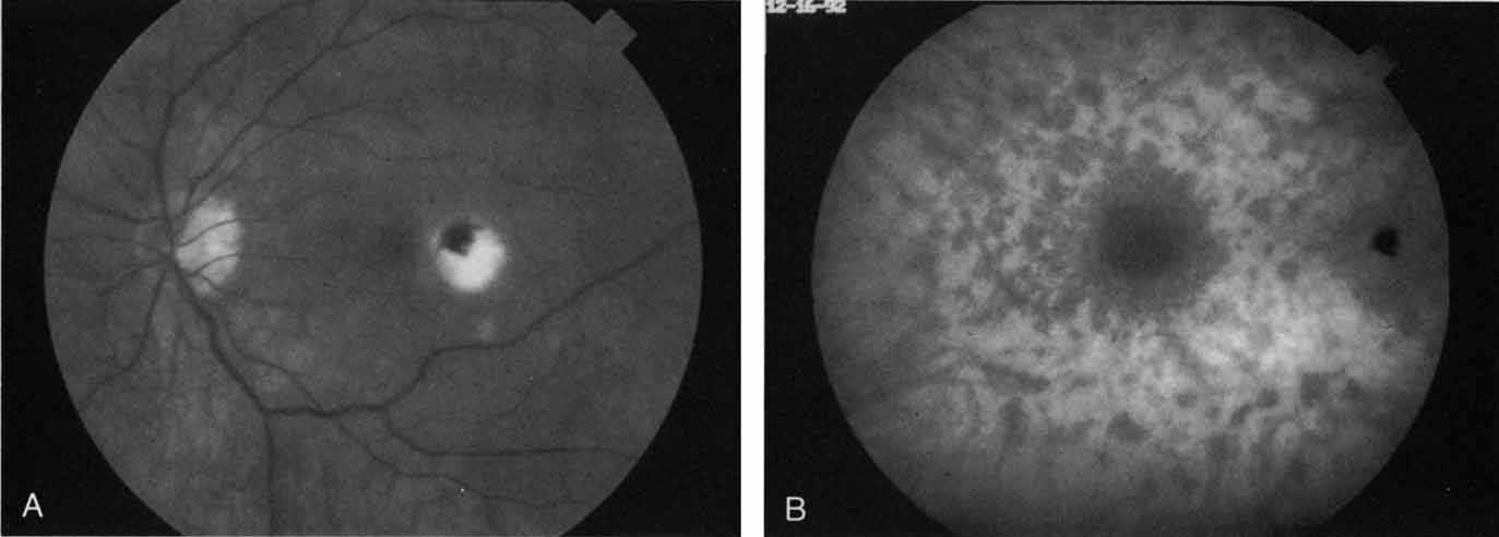
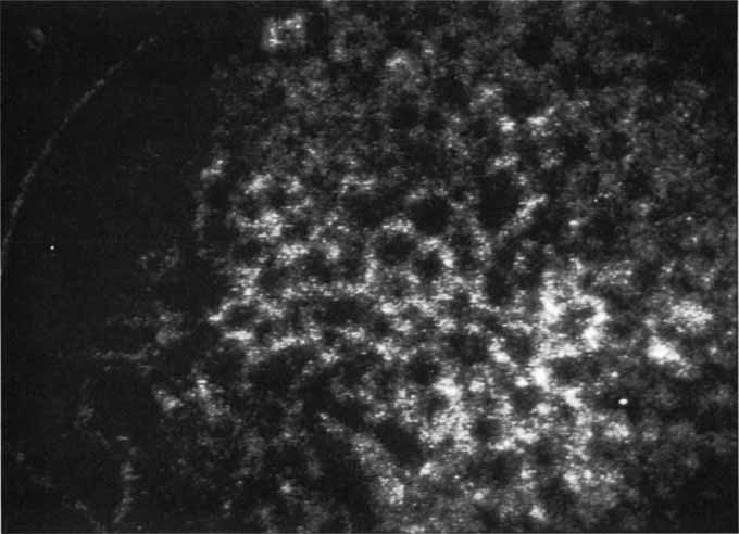
In a landmark article, Yannuzzi and associates18 demonstrated that ICG videoangiography was extremely useful in identifying well-demarcated localized areas of neovascularization in what had been classified as occult CNV by standard fluorescein angiography.19–24 In this study, 39% of 129 patients with “occult CNV” originally diagnosed as determined by fluorescein angiography were given a revised diagnosis of “well-defined neovascular lesions” based on the information obtained from the ICG study (Fig. 1). One study revealed that approximately 40% of patients with occult CNV diagnosed actually presented with early, well-defined focal areas of fluorescence on ICG videoangiography.25 They further defined two potential subgroups of occult CNV: those with and those without serous pigment epithelial detachments (PEDs) accompanying the occult neovascular process. They also pointed out that ICG angiography offered a potential advantage in identifying neovascular lesions when there was clinical evidence of recurrent CNV after previous laser photocoagulation treatment.

Fig. 1 A. Late-phase fluorescein angiogram of a patient with exudative age-related macular degeneration demonstrating diffuse hyperfluorescence and staining of a serous retinal pigment epithelial detachment. No localized area of classic neovascularization was identified, and the patient was classified as having occult choroidal neovascularization. B. Mid-phase indocyanine green angiogram demonstrating a focal spot of hyperfluorescence presumed to represent a localized area of choroidal neovascularization.

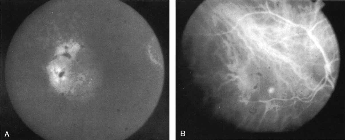
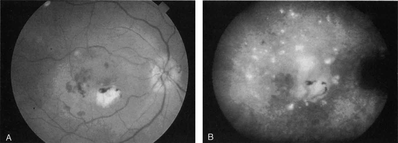
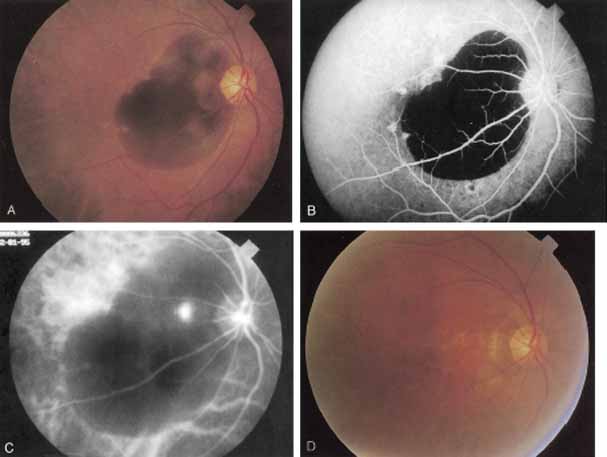
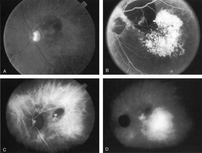
Subsequently, Yannuzzi and colleagues25 and Guyer and associates26 evaluated the usefulness of ICG angiography in identifying localized areas of CNV in patients with occult CNV with or without a serous PED. In a careful evaluation of more than 680 patients, they identified 22% of patients with localized lesions that might be amenable to laser therapy—lesions that would otherwise be classified as untreatable based on guidelines for laser photocoagulation (Figs. 2 and 3). As a result of this improved imaging technique, as many as two to three times the number of patients would have been potentially eligible for laser treatment than would have been treatable based on fluorescein angiography alone.

Fig. 2 A. Late-phase fluorescein study of a patient with exudative age-related macular degeneration demonstrating hyperfluorescence of the serous component of a retinal pigment epithelial detachment. There is relative hypofluorescence on the nasal aspect of the pigment epithelial detachment, and some irregular staining in the superior margin. No well-defined area of neovascularization was identified. B. Mid-phase indocyanine green angiogram demonstrating a localized area of intense hyperfluorescence corresponding to the localized area of active choroidal neovascularization. The pigment epithelial detachment is hypofluorescent. A patient with such findings was considered to be potentially eligible for laser photocoagulation therapy.

Fig. 3 A. Clinical photograph of a patient with subretinal hemorrhage secondary to exudative age-related macular degeneration. B. Late-phase fluorescein study demonstrating hypofluorescence caused by blockage by the subretinal hemorrhage. No area of choroidal neovascularization could be identified. C. Late-phase indocyanine green (ICG) study demonstrating a localized area of intense hyperfluorescence corresponding to active choroidal neovascularization. The thicker areas of subretinal hemorrhage continued to block fluorescence, even in the ICG study, but a localized area of presumed neovascularization is identified that would be potentially amenable to laser treatment.

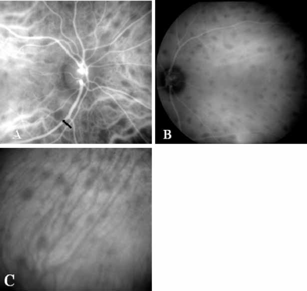
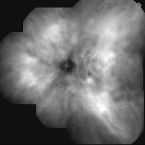
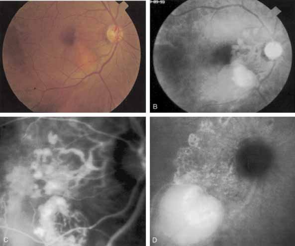
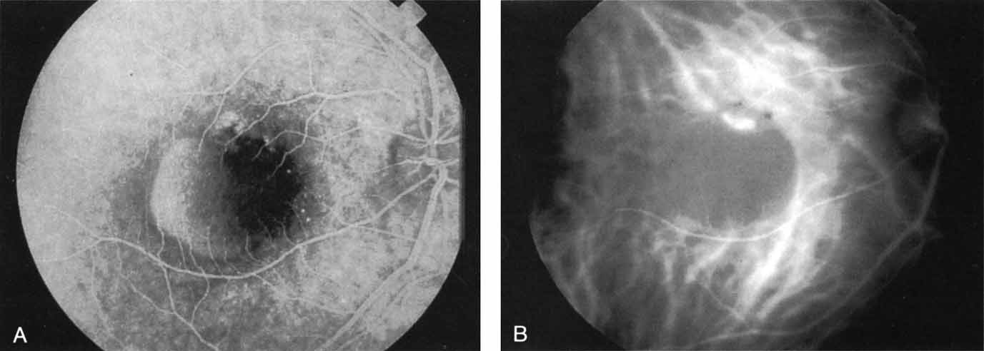
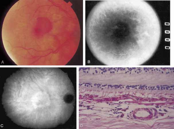
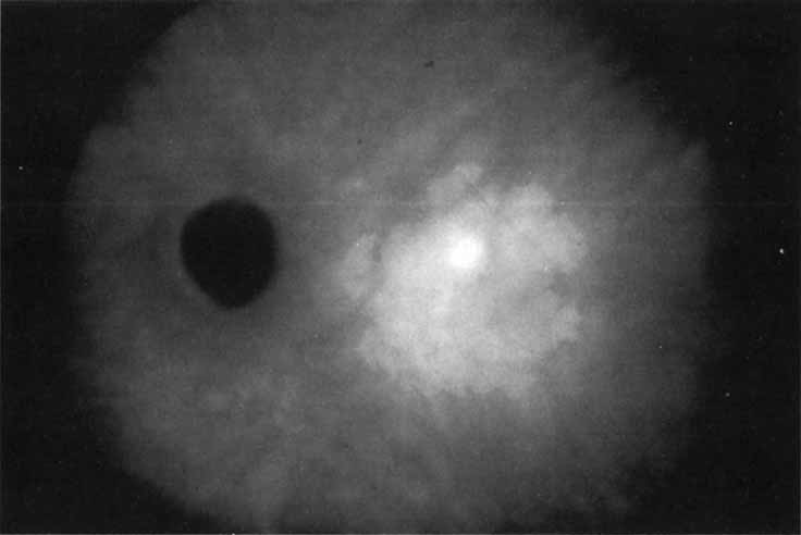
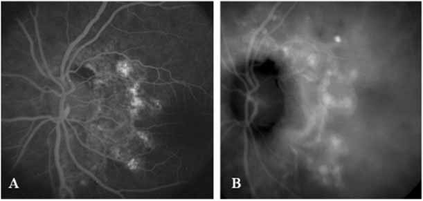
An important report by Chang and co-workers27 lends support to the rationale for interpreting the hyperfluorescence seen on ICG angiography as CNV. In this clinicopathologic study, a patient was identified who had experienced subretinal hemorrhage with early signs of occult CNV on fluorescein angiography (Fig. 4A). Fluorescein angiography demonstrated blocked fluorescence (Fig. 4B). The ICG study, however, demonstrated late staining in a well-circumscribed fashion, which the authors interpreted as a “plaque” of occult CNV (Fig. 4C). When the patient died, this area was evaluated and studied histopathologically and compared with the picture seen on ICG angiography. The area of hyperfluorescence on the ICG study corresponded precisely to a thin layer of fibrovascular tissue beneath the pigment epithelium and neurosensory retina, confirming that the late-staining tissue imaged with ICG angiography was truly a neovascular membrane (Fig. 4D).

Fig. 4 A. Clinical photograph of a patient with exudative age-related macular degeneration demonstrating subretinal hemorrhage. B. Late-phase fluorescein study demonstrating blocked fluorescence from the subretinal hemorrhage. C. Late-phase indocyanine green (ICG) angiogram demonstrating a well-defined area of hyperfluorescence presumed to represent a localized plaque of choroidal neovascularization. Several months after this angiographic study was performed, the patient died and the eyes were obtained for histopathologic examination. D. Hematoxylin and eosin preparation of a section through the retina and choroid in an area corresponding to the hyperfluorescence noted on the ICG study. There is a focal area of fibrovascular tissue beneath the retina. The histopathologic study confirmed the presence of a neovascular membrane in an area corresponding to the zone of hyperfluorescence noted on the ICG study. (Chang TS, Freund KB, De la Cruz Z, et al: Clinicopathologic correlation of choroidal neovascularization demonstrated by indocyanine green angiography in a patient with retention of good vision for almost four years. Retina 14:114, 1994)

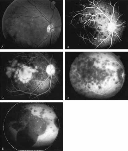
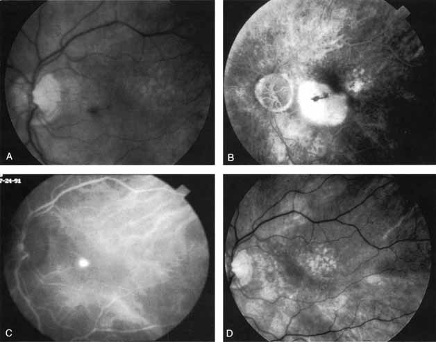
With this clinical diagnostic and histopathologic information available, pilot studies were performed to determine the practicality of using ICG angiographic guidance in the treatment of occult CNV. Slakter and associates28 performed laser photocoagulation treatment on 79 eyes with occult CNV. The occult CNV was successfully eliminated in 57% of patients who underwent ICG-guided treatment (Figs. 5 and 6). The authors found the success rate to be higher (66%) for patients with CNV not associated with PEDs than for those with PEDs (43%). Visual acuity improvement or stabilization was achieved in 57% of all patients. Recurrences were more frequent and more difficult to control in those patients who had associated PEDs on initial clinical presentation. Additional independent studies have reported similar diagnostic and treatment outcomes with the use of ICG angiography in patients with occult CNV.29,30

Fig. 5 A. Clinical photograph of a patient with exudative age-related macular degeneration with secondary subretinal hemorrhage. B. Late-phase fluorescein angiogram demonstrating blocked fluorescence from the subretinal hemorrhage. C. Mid-phase indocyanine green (ICG) study demonstrating blocked fluorescence from the thick areas of subretinal hemorrhage, but a localized area of intense hyperfluorescence representing active choroidal neovascularization was noted in the papillomacular bundle. Laser photocoagulation treatment was applied to this spot only. D. Clinical photograph obtained 3 months later demonstrates an atrophic scar at the site of laser treatment and complete resolution of the subretinal hemorrhage.

Fig. 6 A. Clinical photograph of a patient with age-related macular degeneration demonstrating a small amount of subretinal hemorrhage and a localized retinal pigment epithelial (RPE) detachment. B. Late-phase fluorescein angiogram demonstrating confluent hyperfluorescence of the serous RPE detachment. No localized area of neovascularization that would have been amenable to laser treatment was identified. C. Mid-phase indocyanine green angiogram demonstrating a focal, intensely hyperfluorescent spot representing a localized area of choroidal neovascularization. Laser photocoagulation treatment was applied only to this area. D. Clinical photograph obtained 2 months later demonstrates complete resolution of the exudative changes and a small atrophic scar at the site of laser treatment. (Yannuzzi LA, Slakter JS, Sorenson JA, et al: Digital indocyanine green videoangiography in choroidal neovascularization. Retina 12:191, 1992)

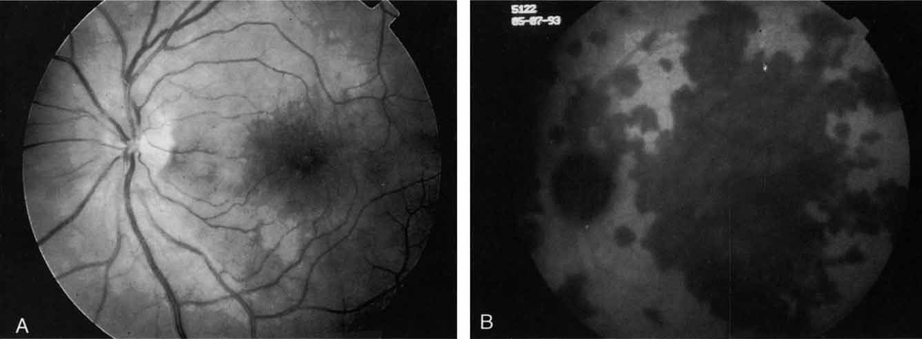
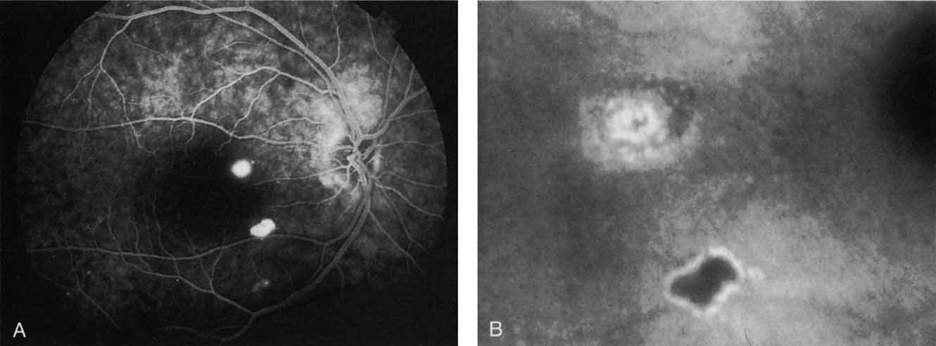
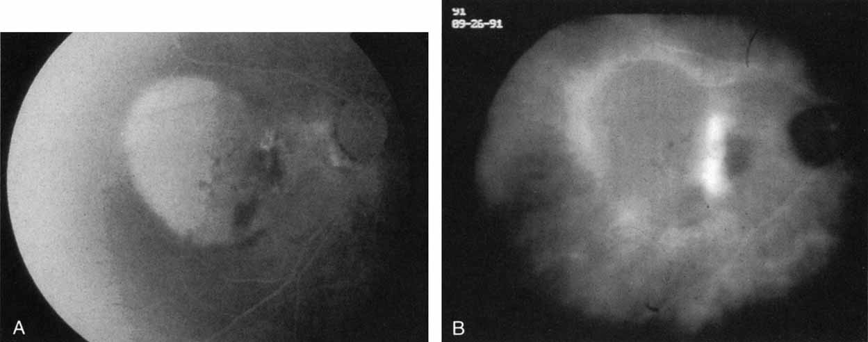
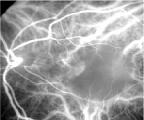
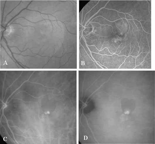
Sorenson and colleagues31 reported on the diagnostic and therapeutic ability of ICG angiography in patients who had clinical signs of recurrent CNV but who were found to have occult membranes on fluorescein angiography. In a group of 66 patients, 97% were identified as having localized areas of hyperfluorescence on ICG angiography consistent with recurrent CNV (Fig. 7). In laser photocoagulation treatment performed in a subgroup of 29 patients, 62% achieved anatomic resolution and stabilization of the exudative process over time. Visual acuity improved in 66%, 45% of whom achieved a visual acuity of 20/100 or better.

Fig. 7 A. Late-phase fluorescein study in a patient with exudative manifestations of recurrent choroidal neovascularization. There is marked hyperfluorescence of the serous component of the retinal pigment epithelial detachment, blocked fluorescence along its nasal margin, and irregular staining in the papillomacular bundle. B. Late-phase indocyanine green study demonstrating a band of intense hyperfluorescence representing recurrent choroidal neovascularization. This is noted along the margin of a localized area of hypofluorescence in the papillomacular bundle, which was the site of the previous photocoagulation treatment. Temporally, a mildly hypofluorescent retinal pigment epithelial elevation is noted. The contrast of the hyperfluorescent active recurrent neovascularization against the relative hypofluorescence of the treatment site and retinal pigment epithelial elevation helps to identify and localize the recurrent process. (Yannuzzi LA, Slakter JS, Sorenson JA et al: Digital indocyanine green videoangiography in choroidal neovascularization. Retina 12:191, 1992)

In essence, the technique of ICG-guided laser treatment is a repetition of traditional principles employed with fluorescein angiographic imaging. The technique involves identification of a staining subretinal lesion imaged with ICG angiography, photocoagulation under ICG guidance, obliteration of the lesion, and resolution of the secondary serosanguineous complications. Obviously, stabilization or improvement of vision is a desired outcome.

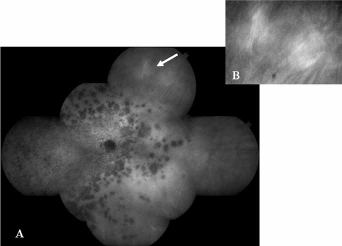
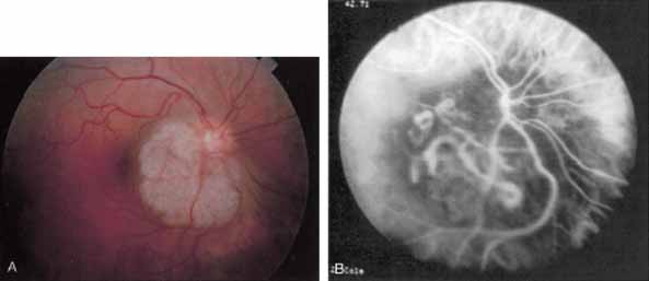
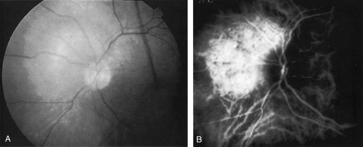
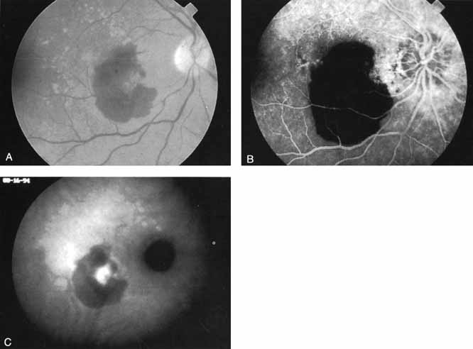
Unlike fluorescein angiography, however, ICG angiographic studies have pointed out that certain principles of laser photocoagulation treatment may not hold with this new imaging capability. This is particularly true with the concept that all areas of CNV need to be obliterated to achieve a successful anatomic result. A careful evaluation of ICG angiograms in some patients with occult CNV has revealed that two forms of neovascular lesions may exist: (1) localized, intensely hyperfluorescent leaking areas of “active” CNV; and (2) more subtle and larger areas of hyperfluorescence with minimal leakage, representing “quiescent” portions of the neovascular complex (Fig. 8). A pilot study by Guyer and colleagues22 demonstrated that localized photocoagulation treatment applied to the active area of CNV alone might result in successful and long-term anatomic stabilization and improvement in the visual acuity in some patients (Fig. 9). A subsequent review of these lesions has demonstrated that they may represent a subpopulation of occult CNV known as polypoidal choroidal neovascularization, described later.

Fig. 8 Late-phase indocyanine green angiogram demonstrating a well-circumscribed are of mild hyperfluorescence in the central macula. Within this region is a localized area of more intense hyperfluorescence. This focal area is presumed to represent a more active site of choroidal neovascularization. The larger area represents a plaque of relatively more quiescent vascular growth.

Fig. 9 A. Clinical photograph of a patient with exudative age-related macular degeneration with evidence of subretinal hemorrhage and shallow neurosensory detachment centrally. B. Late-phase fluorescein study demonstrating blocked fluorescence in the superior macula and intense hyperfluorescence and staining in the area of the neurosensory detachment (A and B, Yannuzzi LA, Slakter JS, Sorenson JA, et al: Digital indocyanine green (ICG) videoangiography and choroidal neovascularization Retina 12:191, 1992). C. Early-phase indocyanine green study demonstrating a focal area of hyperfluorescence beneath the hemorrhage. This area was presumed to be the site of more active neovascularization, D. Later-phase ICG study demonstrating the continued hyperfluorescence of the focal spot in the papillomacular bundle as well as a larger plaque of choroidal neovascularization. Laser photocoagulation was applied only to the more active focal spot of neovascularization.

Flower revealed that feeder vessels (FVs) identified by dynamic ICG angiography, but not by FA, may be amenable to treatment with argon green laser.32 ICG angiography is then performed at regular intervals after treatment to determine the treatment response. A second laser treatment is administered if ICG angiography reveals a patent FV. A 40% success rate was found, initially, with a more recent report of 75% success.33 It appears that dynamic ICG angiography, especially high-speed ICG, detects smaller FVs and can therefore be used in treatment of these vessels. (Fig. 10)

Fig. 10 High-speed indocyanine green angiography image of a feeder vessel.

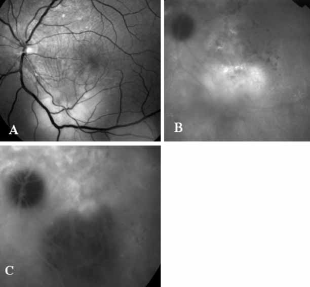
More recently, ICG angiography has begun to play an important role in the management of patients with occult CNV. In particular, the identification and management of two subsets of occult CNV, namely retinal angiomatous proliferation (RAP)34 and polypoidal choroidal neovascularization (PCV),35 are greatly enhanced with ICG angiography. RAP is a distinct subgroup of neovascular AMD in which angiomatous proliferation within the retina is the first manifestation of the process of neovascularization. As the neovascularization proliferates into the outer retina and subretinal space, the angiomatous proliferation is then surrounded by dilated retinal vessels, hemorrhages (preretinal, intraretinal, and subretinal), and exudates. One or more of the related, compensatory retinal vessels perfuse and drain the neovascularization, occasionally forming a retinal–retinal anastomosis (RRA). In these patients, the same indistinct staining seen in occult CNV is present on a fluorescein angiogram. Therefore, most cases of RAP require the use of ICG angiography to make the diagnosis (Fig. 11).34

Fig. 11 A. Clinical photograph of a patient with retinal angiomatous proliferation (RAP). There is a hemorrhage with a retinal pigment epithelial detachment. B. Early-phase fluorescein angiogram demonstrates the retinal–retinal anastomosis. C. Late-phase fluorescein study demonstrates the late-staining hyperfluorescence seen in RAP lesions with a hyperfluourescent retinal pigment epithelial detachment. D. Mid-phase indocyanine green study demonstrates the hyperfluorescent hot-spot overlying the hypofluorescent retinal pigment epithelial detachment. E. Late-phase ICG angiogram demonstrates late hyperfluorescent leakage (CME) into the retina.

ICG angiograms of RAP lesions reveal a focal area of intense hyperfluorescence corresponding to a so-called hot-spot of neovascularization with some late extension of the leakage within the retina caused by intraretinal neovascularization (IRN) within the deep layers of the retina. As the IRN progresses down into the subretinal space, the neovascularization present in the choroid joins the IRN to form a large, neovascular complex. At this stage, clinical and angiographic evidence of a vascularized PED (V-PED) is often present. ICG is the preferred method of imaging a V-PED because the serous component of the PED remains hypofluorescent while the vascular component displays hyperfluorescence. ICG angiography may be able to capture direct communication between the retinal and choroidal component of the neovascular complex as they meet to form a retinal choroidal anastomosis (RCA).34 Treatment options of RAP lesions include: traditional laser photocoagulation of the stage 1 and early stage 2 lesions, surgical excision of stage 2 lesions in conjunction with laser diathermy, as well as PDT, alone and in combination with other treatments, particularly triamcinolone acetonide.36,37 Other therapies, both singly and in combination, are currently undergoing investigation for this unique form of neovascular AMD.

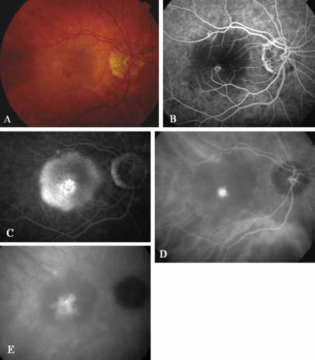
Although initially reported exclusively in middle-aged, black females, PCV has since been recognized as a variant of CNV that can be found in all patients with AMD. This entity is characterized by the presence of an inner choroidal vascular network ending in an aneurysmal bulge clinically seen as a red–orange, spheroid, polyp-like structure. Leakage and bleeding from the choroidal vascular abnormalities result in multiple, recurrent, serosanguinous RPE detachments.25,39,40,41 ICG can be used to identify and characterize the vascular abnormality with high sensitivity and specificity.19–21,40–59 Early-phase images of the lesions show a distinct network of vessels within the choroid. Patients with juxtapapillary involvement show a radial, arching pattern with an inner network of vascular channels extending and connecting with smaller, spanning branches that are more numerous and increasingly prominent at the edge of the PCV lesion.

Larger choroidal vessels in the PCV lesion begin to fill before retinal vessels but appear to fill at a slower rate than the retinal vessels. The lesion initially appears noticeably hypofluorescent relative to the surrounding, uninvolved choroid, but soon after the network is first visible with ICG angiography, small, hyperfluorescent “polyps” can be seen within the choroid. These polypoidal structures correspond to the red–orange choroidal excrescence seen clinically. They leak slowly into the surrounding hypofluorescent area, creating increasing hyperfluorescence. In the late phases of the ICG angiogram, a uniform washout pattern is seen as the dye disappears from the polypoidal vascular structure; the late-staining characteristic of occult CNV is not seen in PCV. PCV may be localized to the macular area without any peripapillary involvement. It may present as a network of small branching vessels ending in polypoidal dilation best-imaged with ICG angiography. (Fig. 12)

Fig. 12 A. Fluorescein study revealing indistinct peripapillary hyperfluorescence and leakage. B. ICG angiogram reveals distinct polypoidal lesions surrounding the disc in this patient with polypoidal choroidal vasculopathy.

ICG angiography has clearly provided new and important diagnostic information, identifying well-defined, potentially laser-treatable lesions in approximately 30% of occult cases.21,30 In addition, the successful elimination of the exudation in patients with occult CNV by obliteration of the hyperfluorescent lesion with laser photocoagulation treatment suggests that ICG-guided laser treatment of occult CNV may be a useful technique in the management of some of these difficult cases (Fig. 13).

Fig. 13 A. Clinical photograph of a patient with pigmentary figures and a surrounding retinal pigment epithelial detachment. B. Fluorescein study reveals an indistinct pattern of hyperfluorescence located just below a serous pigment epithelial detachment. C. Indocyanine green (ICG) angiogram demonstrates a hyperfluorescent small polypoidal vessel at the inferior margin of a hypofluorescent retinal pigment epithelial detachment. D. Late-phase ICG study reveals some late hyperfluorescence corresponding to a small plaque inferior to the focal polypoidal lesion seen in C. In this case, the ICG images were used to guide the treatment of the lesion in this patient. Only the hyperfluorescent spots evident on the early-phase ICG were treated.

Perhaps most importantly, ICG angiography has provided a better overall understanding of the complexity of patients with occult CNV. Certainly this diagnosis may be an oversimplification of the nature of the condition. The presence or absence of a serous PED accompanying this disorder may have a bearing not only on the treatment course but also on the natural history of the disease. In addition, not all areas of CNV may be equivalent: areas that are more actively proliferating may represent a larger threat to visual acuity than the more quiescent plaques of CNV. These larger areas of quiescent vessels may, in fact, be compatible with long-term good visual acuity and may not require therapy. Undoubtedly, much more remains to be learned regarding the nature of these complex lesions, but ICG angiography has clearly been demonstrated to play an important role in the evaluation and management of these patients.

Back to Top
CHOROIDAL INFLAMMATORY DISEASE
The value of ICG angiography appears promising in the area of choroidal inflammatory disease. The enhanced imaging of the pathologic lesions in a number of inflammatory conditions with the ICG technique has led to a better recognition and classification of the disease and may lead to better management options.
Back to Top
BIRDSHOT CHORIORETINOPATHY

In birdshot chorioretinopathy, the typical clinical appearance of ovoid, cream-colored lesions deep in the retina is easily recognizable on examination. Fluorescein angiography frequently lends very little new information, with minimal irregular hypofluorescence because of blockage of the pigment epithelium. With ICG angiography, the uniform background choroidal fluorescence permits the lesions to stand out in high relief. They appear as ovoid, well-demarcated areas of hypofluorescence that appear to be deep in the choroidal layers (Fig. 14). They have a “vasotropic orientation” following along the distribution of the larger choroidal vessels. The dots visualized with ICG are similar in size to, but are more numerous than, those seen clinically.

Fig. 14 A. Mid-phase indocyanine green (ICG) study in a patient with Birdshot choroidopathy. The arrow corresponds to just one of the multiple sites of hypofluorescence caused by the inflammatory lesions in the choroid. B. Late-phase ICG demonstrating multiple hypofluorecent lesions. C. ICG study in the mid-peripheral fundus demonstrating that these hypofluorescent lesions are aligned along the choroidal vessels.

Back to Top
MULTIPLE EVANESCENT WHITE DOT SYNDROME

In multiple evanescent white dot syndrome (MEWDS), the typical fluorescein appearance in the acute phase of the disease involves early-phase hyperfluorescence of dots in a ring formation, corresponding to the clinically evident lesions.60 In general, these dots are found only in the posterior pole. Interestingly, ICG angiography reveals an increased number of lesions beyond those clinically evident. The angiogram initially reveals a stellate pattern of hypofluorescent spots in a ring formation.61 In the later phases of ICG angiography, however, there are more widely distributed areas of hypofluorescent spots, frequently extending far into the periphery (Fig. 15).

Fig. 15 A. Composite montage of the ICG angiogram in a patient with multiple evanescent white dot syndrome (MEWDS). Note the numerous large hypofluorescent spots with overlying smaller hypofluorescent dots. These lesions evident on the ICG study far outnumbered the clinically apparent ones. The arrow demonstrates the area that was enlarged in B. B. An interesting finding on the ICG corresponds to these areas of relative hyperfluorescence of the choroidal vessels, which indicate a localized inflammatory reaction.

The widespread nature of these lesions would help to explain the generalized nature of this condition, which is exemplified by abnormalities frequently detected in electroretinographic testing.62 In addition, some patients with MEWDS have been reported60,63 to have blind-spot enlargement on visual field testing. This was unexplained by either clinical or fluorescein angiographic examination and was attributed to an idiopathic peripapillary retinal dysfunction. ICG angiography in some of these patients has revealed confluent areas of hypofluorescent spots surrounding the optic nerve64 (Fig. 15A). This may provide the first evidence of a region of hypoperfusion or inflammation that may lead to secondary dysfunction and blind spot enlargement. It has been shown that resolution of the enlarged blind spot and return of vision does not correlate completely with the disappearance of hypofluorescent areas on ICG angiography; these lesions often remain visible on ICG study. These findings suggest that MEWDS may result in persistent abnormalities in choroidal circulation, even after vision has been restored.

Back to Top
ACUTE MULTIFOCAL POSTERIOR PLACOID PIGMENT EPITHELIOPATHY

In acute multifocal posterior placoid pigment epitheliopathy, discrete hypofluorescent geographic areas are present both in the posterior pole and extending into the mid-periphery on ICG angiography65,66 (Fig. 16). These hypofluorescent areas are more extensive than noted on either clinical or fluorescein angiographic examination. They are noted in the early phases of the ICG study and persist late, suggesting that ischemic changes in the choroidal circulation occur in this disease. The hypofluorescence remains even once lesions have healed.

Fig. 16 A. Clinical photograph of a patient with acute multifocal placoid pigment epitheliopathy (AMPPE). There is extensive involvement of the central macula by the inflammatory lesions. B. Late-phase indocyanine green angiogram demonstrating extensive confluent but irregular areas of marked hypofluorescence. The hypofluorescence in AMPPE is believed to represent a perfusion abnormality within the choroidal circulation.

Back to Top
SERPIGINOUS CHORIORETINOPATHY
In cases of serpiginous choroidopathy, ICG can be used to delineate the acute phase from the subacute stage as each stage shows a distinct pattern on the angiogram. The acute stage of serpiginous choroidopathy is characterized by generalized hypofluorescence in all phases of the study. In the subacute stage, mid-sized and large choroidal vessels can be visualized within the lesion. Delay in or lack of filling in the smaller choroidal vessels, including the choriocapillaris, yields a generalized hypofluorescence in all phases of the study. The subacute phase produces a more detailed picture than the acute stage, because there is resolution of acute inflammatory changes and associated edema. The late-phase in subacute serpiginous chorioretinopathy reveals well-demarcated lesions that represent choroidal perfusion abnormalities and blockage by inflammatory exudates, along with edema of the RPE and of the outer retina. When serpiginous choroidopathy becomes inactive, atrophy of the RPE and choriocapillaris allows for visualization of the deep choroidal vessels.67 A report by Giovannini and co-workers suggests the presence of occult satellite lesions in the choroid seen on ICG angiography without either clinical or fluorescein angiographic evidence.67 It has been suggested that these quiescent lesions represent occult manifestations of the disease.

ICG angiography is particularly useful to distinguish between serpiginous choroidopathy and other choroidal vascular inflammatory diseases such as Vogt-Koyanagi-Harada Syndrome (VKH), ocular sarcoidosis, ocular tuberculosis, and birdshot choroidopathy. In these conditions, there is diffuse choroidal hyperfluorescence in the late-phases of angiography caused by involvement in the large choroidal vessels.

Back to Top
HARADA DISEASE

Harada disease, an inflammatory condition often affecting both eyes in young patients, is typified on fluorescein angiography by multiple focal areas of hypofluorescence early in the study, followed by late confluent leakage in the later phases (Fig. 17A, 17B, and 17C). With ICG angiography, multiple focal hypofluorescent spots are seen in the early phases, many more than were noted on the fluorescein study64,68 (Fig. 17D). The process extends further than the clinically or angiographically involved areas. The spots vary in size and density but appear to be well circumscribed. In the later phases of the ICG study, there is generalized hypofluorescence, with masking of the background choroidal fluorescence by the neurosensory detachment (Fig. 17E). This masking effect covers a large area, with inferior round margins confirming the gravitating nature of the neurosensory elevation. In addition, focal hyperfluorescent spots can be seen, possibly representing sites of active chorioretinal leakage or inflammation. In one patient, steroid therapy resulted in a marked resolution of clinical findings within 1 week.68 Repeat ICG angiography revealed a marked resolution of the hypofluorescent lesions as well as the hyperfluorescent spots. New areas of hypofluorescence did appear, the significance of which remains undetermined (Fig. 17E).

Fig. 17 Clinical photograph of a patient with Harada disease demonstrating multiple serous elevations of the retinal pigment epithelium with an overlying shallow neurosensory detachment. B. Early-phase fluorescein angiogram demonstrating hypofluorescent spots at the site of localized inflammation. C. Late-phase fluorescein study demonstrating hyperfluorescence in a confluent nature in the central macula. D. Early-phase indocyanine green (ICG) study demonstrating hypofluorescent spots in the central macular region. Note that the lesions are more numerous and more widely distributed than noted on clinical or fluorescein angiographic examination. E. Late-phase ICG study demonstrating extensive areas of confluent hypofluorescence. Note the curvilinear and gravitating nature of these hypofluorescent lesions, which are believed to represent blocked fluorescence from the shallow neurosensory detachment. Focal hyperfluorescent spots are noted within this region, which may represent areas of more active inflammation.

Back to Top
MULTIFOCAL CHOROIDITIS

Most promising in this category of inflammatory choroidopathies is a better detection and understanding of the lesions of multifocal choroiditis. Multiple large, scattered, hypofluorescent spots are seen on ICG angiography, particularly in the later phases of the angiogram69 (Fig. 18). These lesions are not evident either on clinical examination or on fluorescein angiography. In addition to having these larger hypofluorescent lesions, patients with multifocal choroiditis have smaller dot-like lesions and hyperfluorescent foci that do not correlate with lesions seen clinically or by FA. They involve the posterior pole and in some patients extend into the mid-periphery. There is also a “papillotropic” involvement, with confluent hypofluorescent lesions surrounding the optic nerve,69 which may be useful in understanding the associated blind-spot enlargement evident in this condition.70 Other zonal visual field defects have been associated with this condition,70 and corresponding ICG abnormalities have been documented in some patients.69

Fig. 18 A. Clinical photograph of a patient with active inflammation associated with multifocal choroiditis. An atrophic scar from previous laser photocoagulation treatment is noted in the temporal macula. The patient reported visual disturbance, and an enlarged blind spot was noted on visual field testing. B. Late-phase indocyanine green study demonstrating a multitude of hypofluorescent spots that were larger than those seen with multiple evanescent white dot syndrome and more numerous and more extensively distributed than appreciated on clinical examination. Note that there is marked confluence of these lesions around the optic nerve, which may help to explain the enlarged blind spot noted on visual field testing.

Natural history data indicate that with progressive increase in vitritis and associated choroidal inflammation, an increase in the number and extent of these hypofluorescent lesions can be documented.40 Furthermore, use of oral steroid therapy as a means of controlling this inflammatory process has been documented to produce not only clinical resolution of the inflammatory process but also corresponding resolution of the hypofluorescent lesions seen on ICG angiography.69 These clearly demonstrated lesions not only may lead to a better understanding of the process of inflammation involved in multifocal choroiditis and potential management strategies but also may serve to differentiate this condition from the ocular histoplasmosis syndrome.

Back to Top
OCULAR HISTOPLASMOSIS SYNDROME

In contrast to multifocal choroiditis, ocular histoplasmosis syndrome does not demonstrate any large, hypofluorescent lesions on the ICG angiographic examination.69 Instead, there are frequently mid- and late-phase hyperfluorescent lesions in the posterior pole in areas that appear normal both clinically and on fluorescein angiography (Fig. 19). These hyperfluorescent lesions may represent sites of subclinical choroidal inflammation. The presence of these lesions on ICG angiography not only may serve to distinguish the histoplasmosis syndrome but also may serve to explain the apparent de novo appearance of new atrophic spots and CNV in what was previously presumed to be normal chorioretinal tissue.

Fig. 19 A. Clinical photograph of a patient with recurrent choroidal neovascularization associated with ocular histoplasmosis syndrome. A few focal “histo spots” are noted in the superior macula. B. Late-phase indocyanine green study demonstrating hyperfluorescence in the central macula corresponding to recurrent neovascularization. Multiple intensely hyperfluorescent dots are noted throughout the macular region, which do not correspond to any detectable lesion noted clinically or via fluorescein angiography. These focal spots may represent sites of subclinical inflammation at the level of the choroid.

Back to Top
CENTRAL SEROUS CHORIORETINOPATHY

In the early phases of ICG angiography, patients with central serous chorioretinopathy (CSC) demonstrate widespread choroidal vascular hyperpermeability71,72 (Fig. 20). These permeability abnormalities may precede visual symptoms or detectable clinical and fluorescein angiographic changes. Although diffuse leakage from the choriocapillaris is clearly noted on the ICG study, focal active leaks at the level of the retinal pigment epithelium remain better defined on fluorescein angiography.

Fig. 20 Composite photo of a patient with central serous chorioretinopathy. The extensive areas of hyperfluorescence represent areas of presumed choroidal hyperpermeability.

Numerous small, serous PEDs are seen on ICG angiography but may not be detectable with standard fluorescein examination.72 These serous PEDs are hyperfluorescent in the early phases of ICG angiography because of transmission of the fluorescence by the hyperpermeable choroidal vessels. In the late stages of ICG angiography, however, the PEDs become hypofluorescent centrally, with a ring of hyperfluorescence at their margins (Fig. 21). These findings may be caused by accumulation of ICG dye at the margins of the detachment, staining of fibrin, which has a high affinity for the ICG molecule, and blockage by subpigment epithelial fluid.

Fig. 21 A. Late-phase fluorescein study demonstrating hyperfluorescent retinal pigment epithelial detachments in this patient with recent visual symptoms. B. High-magnification of the late-phase of the indocyanine green study, which shows the two pigment epithelial detachments that were hyperfluorescent on the fluorescein angiogram (A). The superior lesion is more active, demonstrating a central hypofluorescence surrounded by a ring of hyperfluorescence, characteristic of the pigment epithelial detachments in patients with central serous chorioretinopathy.

In the chronic or recurrent form of this condition, repeated acute detachments of the neurosensory retina result in atrophic changes and oozing or leakage through the retinal pigment epithelium, with a generalized hyperfluorescent pattern seen on fluorescein angiography.73,74 ICG angiography, in contrast, may reveal only late hypofluorescent serous PEDs in this diffuse, decompensated stage of the disease.72 These characteristic hypofluorescent PEDs, often accompanied by hyperfluorescent rings in the late phases of ICG angiography, appear to be characteristic of central serous chorioretinopathy. The finding of these ring-shaped lesions in the late phases of ICG angiography in an older patient with exudative changes in the macula, but lacking characteristics of age-related macular degeneration such as drusen, might point to the diagnosis of central serous chorioretinopathy.

Recently, Yannuzzi, Slakter, Gross et al performed a pilot study that used ICG angiography to guide the treatment of chronic CSC (CCSC) with photodynamic therapy (PDT)75 (Fig. 22). The results from the pilot study were promising with regard to resolution of both chronic neurosensory retinal detachments documented by optical coherence tomography (OCT) and choroidal hyperpermeablity documented on follow-up ICG angiograms. All patients experienced the aforementioned anatomic improvements and no patients lost vision. In that pilot study, 20 eyes of 15 patients with the diagnosis of CCSC were recruited and treated with PDT. The visual acuity improved two or more lines in six eyes (30%) and remained stable in 14 eyes (70%). All cases had complete resolution of the ICG hyperpermeablity at the site of the treatment, which occurred within a 2- to 6-week interval after PDT. Six eyes experienced improved vision and 14 eyes had stabilization of vision. No eye experienced a visual decline. Patients with visual acuity 20/100 or better were found to have improvement in vision, which was statistically significant by Pearson's correlation. Twelve eyes (60%) had complete anatomic resolution of the fluid in the central macula whereas eight (40%) had partial resolution of the exudative detachment, confirmed by OCT. Five eyes (25%) had residual cystic changes and four (20%) had discernible foveal atrophy as determined by OCT evaluations. These eyes had limited or no improvement in visual acuity.75

Fig. 22 A. Clinical photograph of a patient with chronic central serous chorioretinopathy (CCSC). Note the areas of retinal pigment epithelial atrophy, especially superiorly. There is a large neurosensory retinal detachment with white fibrin along the inferior arcade. B. Late-phase ICG study demonstrating the choroidal hyperpermeability and staining of fibrin. This area was treated in a pilot study using ICG-guided photodynamic therapy with verteporfin. C. ICG study 3 weeks after treatment reveals the characteristic circular hypofluorescence found after PDT treatment. There is a decrease in the choroidal hyperpermeability.

Back to Top
IDIOPATHIC POLYPOIDAL CHOROIDAL VASCULOPATHY

Idiopathic polypoidal choroidal vasculopathy, also known as posterior uveal bleeding syndrome, typically affects darkly pigmented persons who exhibit acute exudative manifestations in the posterior pole, including subretinal hemorrhage and, occasionally, CNV.76,77 ICG angiography facilitates a more definitive diagnosis than fluorescein angiography because it achieves a sharper image of the primary lesion,51,77,78 dilated choroidal vessels terminating in polypoidal or aneurysmal excrescences at the level of the choroid (Fig. 23). These vascular changes account for the secondary exudative and hemorrhagic detachments of the retinal pigment epithelium and neurosensory retina that are characteristic of this disorder. Furthermore, ICG angiography may provide a better means of differentiating the relatively benign aneurysm-like changes at the level of the larger choroidal vessels from CNV, a more devastating secondary manifestation of this chronic disease.79

Fig. 23 A. Clinical photograph of a patient with idiopathic polypoidal choroidal vasculopathy demonstrating the branching lesion at the level of the choroid. There is a more nodular elevation in the inferior macula. A hemorrhagic detachment of the retina is noted temporally in a curvilinear fashion. B. “Green-free” photograph that more vividly demonstrates the branching vascular pattern of this condition. C. High-magnification image of an early-phase indocyanine green (ICG) study reveals filling of the vascular channels at the level of the choroid leading to the polypoidal lesions. Note that the inferior, more elevated lesion, which is nodular on clinical examination, consists of an internal network of branching vessels. D. Late-phase ICG study demonstrates ring-like staining of the small polypoidal lesions. The larger lesion shows intense but nonhomogeneous staining.

Back to Top
CHOROIDAL TUMORS

CHOROIDAL MELANOMA

A number of choroidal tumors have been studied with ICG angiography.80,81 Pigmented choroidal melanomas block ICG fluorescence because of the absorption of the near-infrared light by the melanin-containing lesion. As a result, the choroidal and tumor vasculature cannot be visualized through the dense pigmentation. ICG has failed to provide distinguishing features that might help to differentiate a melanoma from other pigmented lesions, such as nevi or pigmented metastatic lesions; however, when a pigmented choroidal melanoma thickens or develops prominent intrinsic vasculature, ICG angiography reflects this change with an increase in fluorescence in the late phase.82

ICG angiography in amelanotic melanomas reveals variable blockage, depending on the amount of pigmentation present in the lesion. Corkscrew vessels have been identified with ICG angiography, but not with fluorescein angiography, in some patients with amelanotic melanoma80,81 (Fig. 24). The meaning of this vascular pattern currently is unclear, but it may eventually assist in the differentiation of these primary ocular tumors from metastatic lesions.

Fig. 24 A. Clinical photograph of a patient with an amelanotic melanoma adjacent to the optic nerve head. B. Mid-phase indocyanine green angiogram demonstrating corkscrew vessels within the melanoma.

CHOROIDAL HEMANGIOMA

ICG angiography definitely provides a clearer delineation of nonpigmented vascular choroidal tumors than does fluorescein angiography. With choroidal hemangioma, marked progressive hyperfluorescence with intense late staining and leakage is seen on the ICG study82 because of the high vascularity of the lesion.80,81,83,84 (Fig. 25). A speckled pattern within the lesion as well as stellate borders have been noted in some patients.80,81,83 Better visualization of the lesions can be achieved as a result of improved imaging through the serosanguineous retinal elevations typically accompanying the hemangiomas.

Fig. 25 A. Clinical photograph of a patient with a choroidal hemangioma adjacent to the optic nerve. (Courtesy of Dr. Carol Shields.) B. Mid-phase indocyanine green angiogram demonstrating hyperfluorescence and staining of the hemangioma, which has well-defined margins. (Courtesy of Dr. Carol Shields.)

Piccolino developed a technique for studying the vasculature of choroidal hemangiomas that involves performing ICG angiography with artificially increased intraocular pressure.85 The increased intraocular pressure serves to slow choroidal circulation, allowing better delineation of the feeding and draining vessels and of the inner circulation of the tumor.

CHOROIDAL METASTASES

ICG angiography may eventually play a role in distinguishing among various metastatic choroidal lesions. Varying ICG angiographic patterns have been noted depending on the vascularity, pigmentation, and location of the lesion within the eye. In one report,80 breast metastases were noted to demonstrate marked blockage on ICG angiography, whereas metastatic thyroid carcinoma86 and bronchial carcinoid tumors80 demonstrated hyperfluorescence. A metastatic skin melanoma demonstrated blockage on ICG angiography, thus rendering it indistinguishable from primary choroidal melanoma.80

CHOROIDAL OSTEOMA

A pattern of small vessels during early ICG angiography is characteristic of choroidal osteomas.80,81 These tiny vessels are often unidentifiable on fluorescein angiography because of rapid leakage from the incompetent vascular endothelium.87 Late hyperfluorescence has been noted diffusely on ICG angiography, the areas containing bony involvement exhibiting variable blockage throughout the study.

Clearly, certain choroidal tumors (e.g., hemangiomas, osteomas) may exhibit fairly characteristic ICG angiographic patterns; however, these ICG studies do not yet rival indirect ophthalmoscopy, slit-lamp biomicroscopy, and good clinical judgment in the management of choroidal tumors. Much more remains to be determined as experience is gained to identify the exact role that ICG angiography will play in the management of these difficult conditions.

The introduction of SLO in ICG has allowed for in vivo imaging of histologically demonstrated microcirculatory patterns (MCP).88 In some cases, the MCP is tied to the behavior of uveal melanoma; in these cases, visualization of the MCP allows for better characterization of tumor activity.89 It has been demonstrated that the presence of complex MCPs on angiograms in eyes with melanocytic lesions is associated with clinical evidence of lesion growth.90

CHOROIDAL ISCHEMIC DISEASES

ICG angiographic imaging of choroidal ischemic diseases (e.g., in toxemia of pregnancy, Purtscher retinopathy) is limited for two major reasons. The first is the biophysical nature of the large ICG protein-bound molecule, which has some restriction with respect to the perfusion of the choriocapillaris. The second difficulty resides in the limitations of currently available imaging systems. The choriocapillaris still remains too fine and intricate a structure to be directly imaged with these digital systems.

Flower91 used a sequential-image subtraction technique to permit imaging of the choriocapillaris in primates. This technique uses photographs obtained at a rate of 30 frames/sec. Specific frames of the study are subtracted from each other, yielding a differential filling picture. In the very early phases of ICG angiography, choroidal lobules can be imaged, filling and then draining, as the study progresses (Fig. 26).92 This technique is promising for the future ICG angiographic study of choroidal ischemic diseases. For the present, these techniques do not yet have useful clinical applications.

Fig. 26 Indocyanine green photograph obtained through a serial subtraction technique demonstrating the lobular nature of the choriocapillaris in the macular region of this patient. (Courtesy of Dr. Robert Flower.)

Other than the current difficulties with imaging the choriocapillaris, the remaining choroidal vessels are well visualized with ICG angiography. In addition, ICG angiography has the capability of imaging vessels in the retrobulbar space. In a study by Mutoh and colleagues,93 ICG angiography was used to examine the retrobulbar vasculature in patients with pathologic myopia. Posterior ciliary arteries can be seen and studied in areas of posterior staphyloma formation.

Back to Top
CONCLUSIONS
In summary, ICG angiography has provided new and interesting information with respect to a number of choroidal abnormalities. Its impact in the area of choroidal ischemic disease, choroidal tumors, and idiopathic disease is still ancillary to clinical examination and the older diagnostic modalities, like ultrasound. With inflammatory diseases, however, better diagnostic as well as differentiation ability may be provided by ICG angiography. This imaging technique may also lead to a better understanding of the natural course and implications for therapy in these conditions. Most importantly, patients with occult CNV secondary to age-related macular degeneration may derive a significant benefit from ICG imaging as better clinically controlled trials provide us with more definitive information, especially with regard to treatment, in this area. With greater clinical experience and additional clinico-pathologic correlations, ICG angiography promises to provide new information for more specific diagnoses, a better understanding of the pathophysiologic mechanisms, and new opportunities for various treatment strategies for all choroidal diseases.
Back to Top
REFERENCES

1. Macular Photocoagulation Study Group: Argon laser photocoagulation for age-related macular degeneration. Arch Ophthalmol 100:912–918, 1992

2. Macular Photocoagulation Study Group: Krypton laser photocoagulation for neovascularized lesions of age-related macular degeneration. Results of a randomized Clinical Trial. Arch Ophthalmol 108:816–824, 1990

3. Macular Photocoagulation Study Group: Argon laser photocoagulation of neovascular maculopathy. Arch Ophthalmol 109:1109–1114, 1991

4. Macular Photocoagulation Study Group: Subfoveal neovascular lesions in AMD: guidelines for evaluation and treatment. Arch Ophthalmol 109:1242–1257, 1991

5. Freund KB, Yannuzzi LA, Sorenson JA: Age-related macular degeneration and choroidal neovascularization. Am J Ophthalmol 115:786–791, 1993

6. Bressler NM: Treatment of Age-Related Macular Degeneration with Photodynamic Therapy (TAP) Study Group. Photodynamic therapy of subfoveal choroidal neovascularization in age-related macular degeneration with verteporfin: one-year results of 2 randomized clinical trials-tap report 1. Arch Ophthalmol 117:1329–1345, 1999

7. Verteporfin in Photodynamic Therapy Study Group: Photodynamic therapy of subfoveal choroidal neovascularization in pathologic myopia with verteporfin. 1-year results of a randomized clinical trial–VIP report no. 1. Ophthalmology 108:841–852.mm, 2001

8. Verteporfin In Photodynamic Therapy Study Group: Verteporfin therapy of subfoveal choroidal neovascularization in age-related macular degeneration: two-year results of a randomized clinical trial including lesions with occult with no classic choroidal neovascularization–verteporfin in photodynamic therapy report 2. Am J Ophthalmol 131(5):541–560, 2001

9. Bressler NM: Treatment of Age-Related Macular Degeneration with Photodynamic Therapy (TAP) Study Group. Photodynamic therapy of subfoveal choroidal neovascularization in age-related macular degeneration with verteporfin: two-year results of 2 randomized clinical trials-tap report 2. Arch Ophthalmol 119:198–207, 2001

10. Orth D, Patz A, Flower RW: Potential clinical applications of indocyanine green choriodal angiography – preliminary report. Eye Ear Nose Throat Mon 55:15–28, 1976

11. Bischoff PM, Flower RW: Ten years experience with choroidal angiography using indocyanine green dye: a new routine examination or an epilogue? Doc Ophthalmol 60:235–291, 1985

12. Hayashi K, DeLaey JJ: Indocyanine green angiography of neovascular membranes. Ophthalmologica 190:30–39, 1985

13. Hayashi K, Hasegawa Y, Tazawa Y, DeLaey JJ: Clinical application of indocyanine angiography to choroidal neovascularization. Jpn J Ophthalmol 33:57–65, 1989

14. Destro M, Puliafito CA: Indocyanine green videoangiography of choroidal neovascularization. Ophthalmology 96:846–853, 1989

15. Guyer DR, Puliafito CP, Mones JM et al: Digital indocyanine green angiography in chorioretinal disorders. Ophthalmology 99:287–291, 1992

16. Scheider A, Schroedel C: High resolution indocyanine green angiography with scanning laser ophthalmoscope. Am J Ophthalmol 108:458–459, 1989

17. Scheider A, Kaboth A, Neuhauser L: Detection of subretinal neovascularization membranes with indocyanine green and infrared scanning laser ophthalmoscope. Am J Ophthalmol 113:45–51, 1992

18. Yannuzzi LA, Slakter JS, Sorenson JA, et al: Digital indocyanine green videoangiography and choroidal neovascularization. Retina 12:191–223, 1992

19. Guyer DR, Yannuzzi LA, Slakter JS, et al: Classification of choroidal neovascularization by digital indocyanine green videoangiography. Ophthalmology 103:2054–2060, 1996

20. Chang B, Yannuzzi LA, Ladas ID, et al: Choroidal neovascularization in second eyes of patients with unilateral exudative age-related macular degeneration. Ophthalmology 102:1380–1386, 1995

21. Destro M, Puliafito CA: Indocyanine green videoangiography of choroidal neovascularization. Ophthalmology 96:846–853, 1988

22. Guyer DR, Yannuzzi LA, Ladas I, et al: Indocyanine green guided laser photocoagulation of focal spots at the edge of plaques of choroidal neovascularization: a pilot study. Arch Ophthalmol 114:693–697, 1996

23. Guyer, DR, Puliafito, CP, Mones, JM, et al: Digital indocyanine green angiography in chorioretinal disorders. Ophthalmology 99:287–290, 1992

24. Lee BL, Lim JI, Grossniklaus HE: Clinicopathologic features of indocyanine green angiography-imaged, surgically excised choroidal neovascular membranes. Retina 16:64–69, 1996

25. Yannuzzi LA, Hope-Ross M, Slakter JS, et al: Analysis of vascularized pigment epithelium detachments using indocyanine green videoangiography. Retina 14:99–113, 1994

26. Yannuzzi LA, Hope-Ross M, Slakter JS, et al: Analysis of vascularized pigment epithelial detachment using indocyanine green videoangiography. Retina 14:99–113, 1994

27. Guyer DR, Yannuzzi LA, Slakter JS, et al: Digital indocyanine green videoangiography of occult-choroidal neovascularization. Ophthalmology 101:1727–1738, 1994

28. Chang TS, Freund KB, De La Cruz Z, et al: Clinicopathologic correlation of choroidal neovascularization demonstrated by indocyanine green angiography in a patient with retention of good vision for almost four years. Retina 14:114–124, 1994

29. Slakter JS, Yannuzzi LA, Sorenson JA, et al: A pilot study of indocyanine-green videoangiography-guided laser treatment of occult-choroidal neovascularization in age-related macular degeneration. Arch Ophthalmol 112:465–472, 1994

30. Regillo SD, Benson WE, Maguire JI, Annesley WH: Indocyanine green angiography and occult choroidal neovascularization. Ophthalmology 101:280–288, 1994

31. Borrillo LJ, Sivalingam A, Martidis A, Federman JL: Surgical ablation of retinal angiomatous proliferation. Arch Ophthalmol 121:558–561, 2003

32. Spaide RF, Sorenson J, Maranan L: Combined photodynamic therapy with verteportin and intravitreal triamcinolone acetonide for choroidal neovascularization. Ophthalmol 110:1517–1525, 2003

33. Lim SI, Sternberg P, Capone A, et al: Selective use of indocyanine green angiography for occult choroidal neovascularization. Am J Ophthalmol 120:75–82, 1995

34. Staurenghi G, Orzalesi N, La Capria A, Aschero M: Laser treatment of feeder vessels in subfoveal choroidal neovascular membranes: a revisitation using dynamic indocyanine green angiography. Ophthalmology 105:2297–2305, 1998

35. Sorenson JA, Yannuzzi LA, Slakter JS, et al: A pilot study of indocyanine-green videoangiography-guided laser treatment of recurrent occult-choroidal neovascularization in age-related macular degeneration. Arch Ophthalmol 112:473–479, 1994

36. Flower RW: Optimizing treatment of choroidal neovascularization feeder vessels associated with age-related macular degeneration. Am J Ophthalmol 134:228–239, 2002

37. Yannuzzi LA, Negrao S, Iida T, et al: Retinal angiomatous proliferation in age-related macular degeneration. Retina 21:416–434, 2001

38. Moorthy RS, Lyon AT, Rabb MF, et al: Idiopathic Polypoidal choroidal vasculopathy of the macula. Ophthalmology 105:1380–1385, 1998

39. Webb RH, Hughes GW, Delori FC: Confocal scanning laser ophthalmoscope. Applied Optics 26:1492–1499, 1987

40. Macular Photocoagulation Study Group: Occult choroidal neovascularization. Influence on visual outcome in patients with age-related macular degeneration. Arch Ophthalmol 114:400–412, 1996

41. Ahuja RM, Stanga PE, Vingerling JR, et al: Polypoidal choroidal vasculopathy in exudative and hemorrhagic pigment epithelial detachments. Br J Ophthalmol 84:479–484, 2000

42. Capone AJr , Wallace RT, Meredith TA: Symptomatic choroidal neovascularization in blacks. Arch Ophthalmol 112:1091–1097, 1994

43. Kwok AKH, Lai TYY, Chan CWN, et al: Polypoidal choroidal vasculopathy in Chinese patients. Br J Ophthalmol 86:892–897, 2002

44. Schneider U, Gelisken F, Inhoffen W: Clinical characteristics of idiopathic polypoidal choroid vasculopathy. Ophthalmologe 98:1186–1191, 2001

45. Shiraga F, Matsuo T, Yokoe S, et al: Surgical treatment of submacular hemorrhage associated with idiopathic polypoidal choroidal vasculopathy. Am J Ophthalmol 128:147–154, 1999

46. Smith RE, Wise K, Kingsley RM: Idiopathic polypoidal choroidal vasculopathy and sickle cell retinopathy. Am J Ophthalmol 129:544–546, 2000

47. Scassellati-Sforzolini B, Mariotti C, Bryan R, et al: Polypoidal choroidal vasculopathy in Italy. Retina 21:121–125, 2001

48. Tateiwa H, Kuroiwa S, Gaun S, et al: Polypoidal choroidal vasculopathy with large vascular network. Graefe's Arch Clin Exp Ophthalmol 240:354–361, 2002

49. Kleiner RC, Brucker AJ, Johnston RL: The posterior uveal bleeding syndrome. Retina 10:9–17, 1990

50. Lafaut BA, Aisenbrey S, van den Broecke C, et al: Polypoidal choroidal vasculopathy pattern in age-related macular degeneration. Retina 20:650–654, 2000

51. Lafaut BA, Leyes AM, Snyers B, et al: Polypoidal choroidal vasculopathy in Caucasians. Graefes Arch Clin Exp Ophthalmol 238:752–759, 2000

52. Lip PL, Hope-Ross MW, Gibson JM: Idiopathic polypoidal choroidal vasculopathy: a disease with diverse clinical spectrum and systemic associations. Eye 5:695–700, 2000

53. Lois N: Idiopathic polypoidal choroidal vasculopathy in a patient with atrophic age-related macular degeneration. Br J Ophthalmol 85:1011–1012, 2001

54. Mohand-Said M, Nodarian M, Salvanet-Bouccara A: Idiopathic polypoidal choroidal vasculopathy: 2 case report. J Franc Ophtalmol 25:517–521

55. Ross RD, Gitter KA, Cohen G, et al: Idiopathic polypoidal choroidal vasculopathy associated with retinal arterial macroaneurysm and hypertensive retinopathy. Retina 16:105–111, 1996

56. Spaide RF, Yannuzzi LA, Slakter JS, et al: Indocyanine green videoangiography of idiopathic polypoidal choroidal vasculopathy. Retina 15:100–110, 1995

57. Yannuzzi LA, Ciardella AP, Spaide RF, et al: The expanding clinical spectrum of idiopathic polypoidal choroidal vasculopathy. Arch Ophthalmol 115:478–485, 1999

58. Yannuzzi LA, Freund KB, Goldbaum M, et al: Polypoidal choroidal vasculopathy masquerading as central serous chorioretinopathy. Ophthalmology 107:767–777, 2000

59. Yannuzzi LA, Sorenson JS, Spaide RF, et al: Idiopathic polypoidal choroidal vasculopathy. Retina 10:1–8, 1990

60. Jampol LM, Sieving PA, Pugh D, et al: Multiple evanescent white dot syndrome: I. Clinical findings. Arch Ophthalmol 102:671–674, 1984

61. Ie D, Glaser BM, Murphy RP et al: Indocyanine green angiography in multiple evanescent white-dot syndrome. Am J Ophthalmol 117:7–12, 1994

62. Sieving PA, Fishman GA, Jampol LM, Pugh D: Multiple evanescent white dot syndrome: II. Electrophysiology of the photoreceptors during retinal pigment epithelial disease. Arch Ophthalmol 102:675–679, 1984

63. Dodwell DG, Jampol LM, Rosenberg M, et al: Optic nerve involvement associated with the multiple evanescent white dot syndrome. Ophthalmology 97:862–868, 1990

64. Slakter JS: Indocyanine green angiography of inflammatory disorders. Presented at the Macula Society Meeting, Palm Desert, CA, February 25, 1994

65. Park D, Schatz H, McDonald HR, Johnson RN: Indocyanine green angiography of acute multifocal posterior placoid pigment epitheliopathy. Ophthalmol 102:1877–1883, 1995

66. Howe LJ, Woon H, Graham EM et al: Choroidal hypoperfusion in acute posterior multifocal placoid pigment epitheliopathy: an indocyanine green angiographic study. Ophthalmology 102:790–798, 1995

67. Giovannini A, Ripa E, Scassellati-Sforzolini B, et al: Indocyanine green angiography in serpiginous choroidopathy. Eur J Ophthalmol 6:299–306, 1996

68. Freund KB: Indocyanine green angiography in Harada's disease. In Flower RW, Yannuzzi LA, Slakter JS (eds): Indocyanine green angiography. St. Louis: CV Mosby, 1997

69. Slakter JS, Giovannini A, Yannuzzi LA, et al: Unpublished data, 1996

70. Khorram KD, Jampol LM, Rosenberg MA: Blind spot enlargement as a manifestation of multifocal choroiditis. Arch Ophthalmol 109:1403–1407, 1991

71. Hayashi K, Hasegawa Y, Tokoro T: Indocyanine green angiography of central serous chorioretinopathy. Int Ophthalmol 9:371–374, 1986

72. Guyer DR, Yannuzzi LA, Slakter JA, et al: Digital indocyanine green videoangiography of central serous chorioretinopathy. Arch Ophthalmol 112:1057–1062, 1994

73. Yannuzzi LA, Shakin JL, Fisher YL, Altomonte M: Peripheral retinal detachments and retinal pigment epithelial atrophic tracts secondary to central serous pigment epitheliopathy. Ophthalmology 91:1554–1572, 1984

74. Yannuzzi LA, Slakter JS, Kaufman SR, Guptka K: Laser treatment of diffuse retinal pigment epitheliopathy. Eur J Ophthalmol 2:103–114, 1992

75. Yannuzzi LA, Slakter JS, Gross NE, et al: Indocyanine green angiography-guided photodynamic therapy for treatment of chronic central serous chorioretinopathy: a pilot study. Retina 23:288–298, 2003

76. Kleiner RC, Brucker AJ, Johnston RL: Posterior uveal bleeding syndrome. Retina 10:9–17, 1990

77. Yannuzzi LA, Wong DW, Sforzolini SB, et al: Polypoidal choroidal vasculopathy and neovascularized age-related macular degeneration. Arch Ophthalmol 17:1503–1510, 1999

78. Pauleikhoff D, Loffert D, Spital G, et al: Pigment epithelial detachment in the elderly. Clinical differentiation, natural course and pathogenetic implications. Graefe's Arch Clin Exp Ophthalmol 240:533–538, 2002

79. Guyer DR, Gragoudas ES, Yannuzzi LA et al: Digital indocyanine green angiography of intraocular tumors. Semin Ophthalmol 8:224–229, 1993

80. Yannuzzi LA, Freund KB, Goldbaum M, et al: Polypoidal choroidal vasculopathy masquerading as central serous chorioretinopathy. Ophthalmol 107:767–777, 2000

81. Shields C, Shields J: Indocyanine green angiography of choroidal tumors. In Flower DW, Yannuzzi LA, Slakter JS (eds): Indocyanine Green Angiography. St. Louis: CV Mosby, 1997

82. Shields CL, Shields JA, De Potter P: Patterns of indocyanine green videoangiography of choroidal tumors. Br J Ophthalmol 79:237–245, 1995

83. Bonnet M, Habozit F, Magnard G: Valeur de l'angiographie en infra-rouge au vert d'indocyanine dans le diagnostic clinique des angiomes de la choroide. Bull Soc Ophthalmol Fr 76:713–716, 1976

84. Quentel G, Coscas G: Angiographie en fluorescence infrarouge au vert d'indocyanine. Bull Soc Ophtalmol Fr 84:559–563, 1984

85. Piccolino FC, Borgia L, Zinicola E: Indocyanine green angiography of circumscribed choroidal hemangiomas. Retina 16:19–28, 1996

86. Bacin F, Buffet JM, Nutel N: Angiographie par absorption, en infrarouge, au vert l'indocyanine: aspects sous le sujet normal et dans les tumeurs choroidennes. Bull Soc Ophtalmol Fr 81:815–819, 1981

87. Kardmas EF, Weiter JJ: Choroidal osteoma. Int Ophthalmol 37:171–182, 1987

88. Singh AD, De Potter P, Shields CL, Shields JA: Indocyanine green angiography and ultrasonography of a varix of the vortex vein. Arch Ophthalmol 100:1283–1284, 1993

89. Seregard S, Spanberg B, Juul C, et al: Prognostic accuracy of the mean of the largest nucleoli, vascular pattens, and PC-10 in posterior uveal melanoma. Ophthalmology 105:485–491, 1998

90. Mueller AJ, Freeman WR, Schaller UC, et al: Complex microcirculation patterns detected by confocal indocyanine green angiography predict time to growth of small choroidal melanocytic tumors. Ophthalmology 109:2207–2214, 2002

91. Flower RW, Klein GJ: Pulsatile flow in the choroidal circulation: a preliminary investigation. Eye 4:310–318, 1990

92. Flower R: Extraction of choriocapillaris hemodynamic data from ICG fluorescence angiograms. Invest Ophthalmol Vis Sci 34:2720–2729, 1993

93. Mutoh T, Sakurai M, Tamai M: Indocyanine green fundus angiography of retrobulbar vasculature. Arch Ophthalmol 113:631–633, 1995

Back to Top