|
|
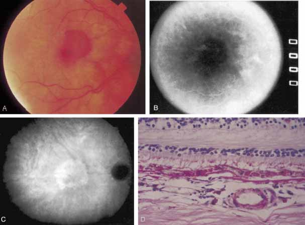
| Fig. 4 A. Clinical photograph of a patient with exudative age-related macular degeneration demonstrating subretinal hemorrhage. B. Late-phase fluorescein study demonstrating blocked fluorescence from the subretinal hemorrhage. C. Late-phase indocyanine green (ICG) angiogram demonstrating a well-defined area of hyperfluorescence presumed to represent a localized plaque of choroidal neovascularization. Several months after this angiographic study was performed, the patient died and the eyes were obtained for histopathologic examination. D. Hematoxylin and eosin preparation of a section through the retina and choroid in an area corresponding to the hyperfluorescence noted on the ICG study. There is a focal area of fibrovascular tissue beneath the retina. The histopathologic study confirmed the presence of a neovascular membrane in an area corresponding to the zone of hyperfluorescence noted on the ICG study. (Chang TS, Freund KB, De la Cruz Z, et al: Clinicopathologic correlation of choroidal neovascularization demonstrated by indocyanine green angiography in a patient with retention of good vision for almost four years. Retina 14:114, 1994) |