|
|
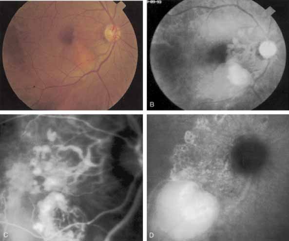
| Fig. 23 A. Clinical photograph of a patient with idiopathic polypoidal choroidal vasculopathy demonstrating the branching lesion at the level of the choroid. There is a more nodular elevation in the inferior macula. A hemorrhagic detachment of the retina is noted temporally in a curvilinear fashion. B. “Green-free” photograph that more vividly demonstrates the branching vascular pattern of this condition. C. High-magnification image of an early-phase indocyanine green (ICG) study reveals filling of the vascular channels at the level of the choroid leading to the polypoidal lesions. Note that the inferior, more elevated lesion, which is nodular on clinical examination, consists of an internal network of branching vessels. D. Late-phase ICG study demonstrates ring-like staining of the small polypoidal lesions. The larger lesion shows intense but nonhomogeneous staining. |