|
|
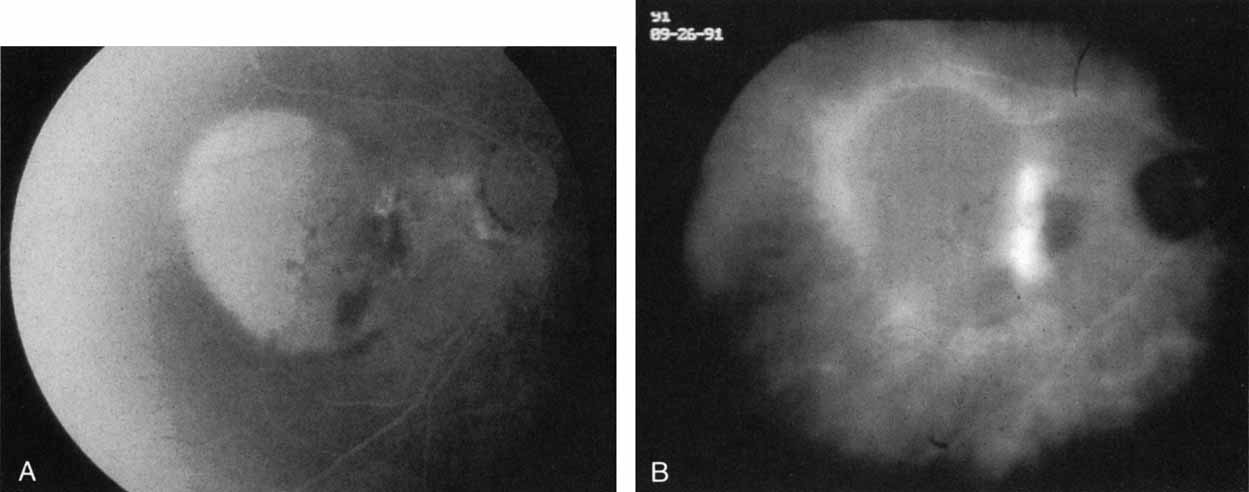
| Fig. 7 A. Late-phase fluorescein study in a patient with exudative manifestations of recurrent choroidal neovascularization. There is marked hyperfluorescence of the serous component of the retinal pigment epithelial detachment, blocked fluorescence along its nasal margin, and irregular staining in the papillomacular bundle. B. Late-phase indocyanine green study demonstrating a band of intense hyperfluorescence representing recurrent choroidal neovascularization. This is noted along the margin of a localized area of hypofluorescence in the papillomacular bundle, which was the site of the previous photocoagulation treatment. Temporally, a mildly hypofluorescent retinal pigment epithelial elevation is noted. The contrast of the hyperfluorescent active recurrent neovascularization against the relative hypofluorescence of the treatment site and retinal pigment epithelial elevation helps to identify and localize the recurrent process. (Yannuzzi LA, Slakter JS, Sorenson JA et al: Digital indocyanine green videoangiography in choroidal neovascularization. Retina 12:191, 1992) |