|
|
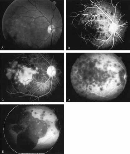
| Fig. 17 Clinical photograph of a patient with Harada disease demonstrating multiple serous elevations of the retinal pigment epithelium with an overlying shallow neurosensory detachment. B. Early-phase fluorescein angiogram demonstrating hypofluorescent spots at the site of localized inflammation. C. Late-phase fluorescein study demonstrating hyperfluorescence in a confluent nature in the central macula. D. Early-phase indocyanine green (ICG) study demonstrating hypofluorescent spots in the central macular region. Note that the lesions are more numerous and more widely distributed than noted on clinical or fluorescein angiographic examination. E. Late-phase ICG study demonstrating extensive areas of confluent hypofluorescence. Note the curvilinear and gravitating nature of these hypofluorescent lesions, which are believed to represent blocked fluorescence from the shallow neurosensory detachment. Focal hyperfluorescent spots are noted within this region, which may represent areas of more active inflammation. |