|
|
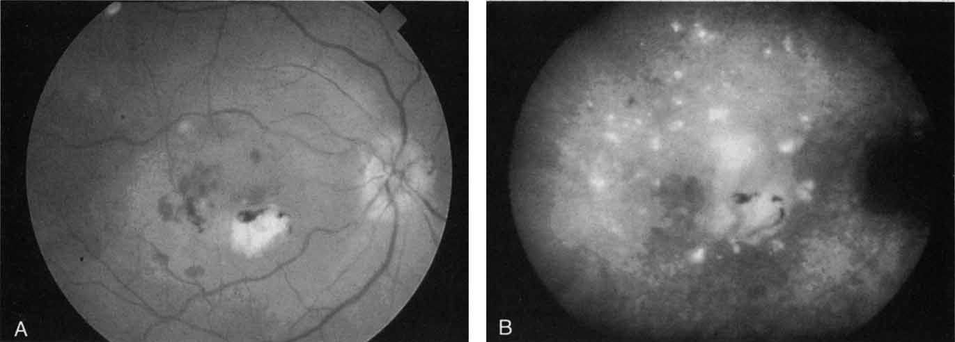
| Fig. 19 A. Clinical photograph of a patient with recurrent choroidal neovascularization associated with ocular histoplasmosis syndrome. A few focal “histo spots” are noted in the superior macula. B. Late-phase indocyanine green study demonstrating hyperfluorescence in the central macula corresponding to recurrent neovascularization. Multiple intensely hyperfluorescent dots are noted throughout the macular region, which do not correspond to any detectable lesion noted clinically or via fluorescein angiography. These focal spots may represent sites of subclinical inflammation at the level of the choroid. |