|
|
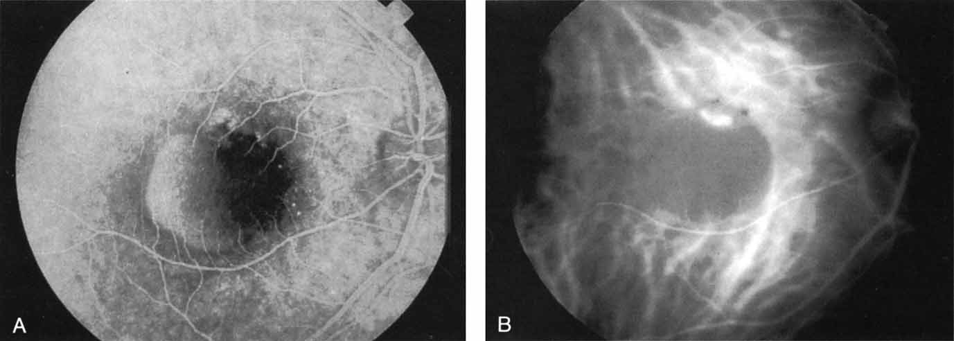
| Fig. 2 A. Late-phase fluorescein study of a patient with exudative age-related macular degeneration demonstrating hyperfluorescence of the serous component of a retinal pigment epithelial detachment. There is relative hypofluorescence on the nasal aspect of the pigment epithelial detachment, and some irregular staining in the superior margin. No well-defined area of neovascularization was identified. B. Mid-phase indocyanine green angiogram demonstrating a localized area of intense hyperfluorescence corresponding to the localized area of active choroidal neovascularization. The pigment epithelial detachment is hypofluorescent. A patient with such findings was considered to be potentially eligible for laser photocoagulation therapy. |