|
|
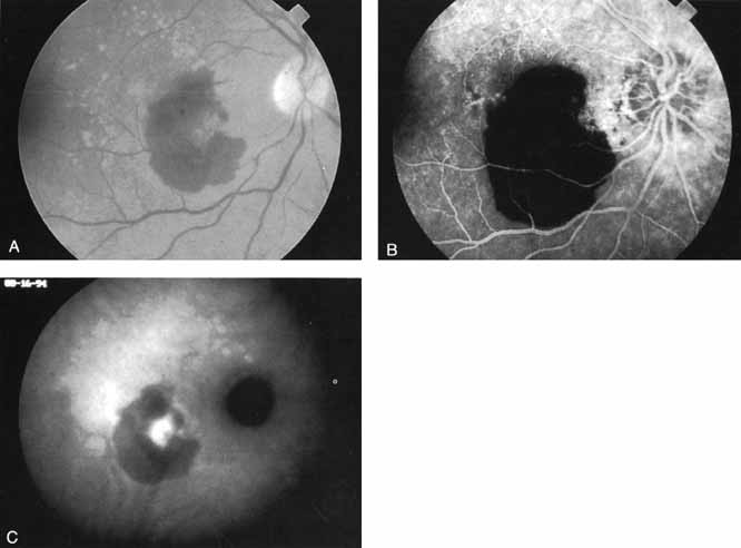
| Fig. 3 A. Clinical photograph of a patient with subretinal hemorrhage secondary to exudative age-related macular degeneration. B. Late-phase fluorescein study demonstrating hypofluorescence caused by blockage by the subretinal hemorrhage. No area of choroidal neovascularization could be identified. C. Late-phase indocyanine green (ICG) study demonstrating a localized area of intense hyperfluorescence corresponding to active choroidal neovascularization. The thicker areas of subretinal hemorrhage continued to block fluorescence, even in the ICG study, but a localized area of presumed neovascularization is identified that would be potentially amenable to laser treatment. |