|
|
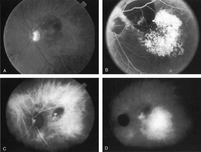
| Fig. 9 A. Clinical photograph of a patient with exudative age-related macular degeneration with evidence of subretinal hemorrhage and shallow neurosensory detachment centrally. B. Late-phase fluorescein study demonstrating blocked fluorescence in the superior macula and intense hyperfluorescence and staining in the area of the neurosensory detachment (A and B, Yannuzzi LA, Slakter JS, Sorenson JA, et al: Digital indocyanine green (ICG) videoangiography and choroidal neovascularization Retina 12:191, 1992). C. Early-phase indocyanine green study demonstrating a focal area of hyperfluorescence beneath the hemorrhage. This area was presumed to be the site of more active neovascularization, D. Later-phase ICG study demonstrating the continued hyperfluorescence of the focal spot in the papillomacular bundle as well as a larger plaque of choroidal neovascularization. Laser photocoagulation was applied only to the more active focal spot of neovascularization. |