|
|
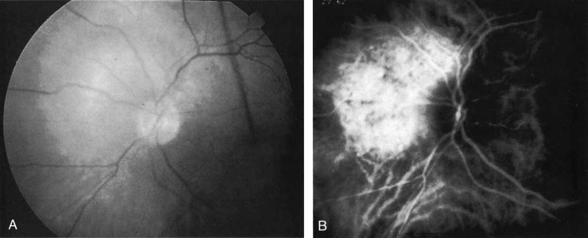
| Fig. 25 A. Clinical photograph of a patient with a choroidal hemangioma adjacent to the optic nerve. (Courtesy of Dr. Carol Shields.) B. Mid-phase indocyanine green angiogram demonstrating hyperfluorescence and staining of the hemangioma, which has well-defined margins. (Courtesy of Dr. Carol Shields.) |