|
|
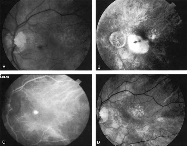
| Fig. 6 A. Clinical photograph of a patient with age-related macular degeneration demonstrating a small amount of subretinal hemorrhage and a localized retinal pigment epithelial (RPE) detachment. B. Late-phase fluorescein angiogram demonstrating confluent hyperfluorescence of the serous RPE detachment. No localized area of neovascularization that would have been amenable to laser treatment was identified. C. Mid-phase indocyanine green angiogram demonstrating a focal, intensely hyperfluorescent spot representing a localized area of choroidal neovascularization. Laser photocoagulation treatment was applied only to this area. D. Clinical photograph obtained 2 months later demonstrates complete resolution of the exudative changes and a small atrophic scar at the site of laser treatment. (Yannuzzi LA, Slakter JS, Sorenson JA, et al: Digital indocyanine green videoangiography in choroidal neovascularization. Retina 12:191, 1992) |