|
|
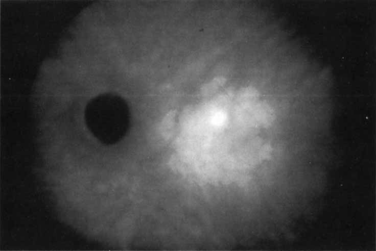
| Fig. 8 Late-phase indocyanine green angiogram demonstrating a well-circumscribed are of mild hyperfluorescence in the central macula. Within this region is a localized area of more intense hyperfluorescence. This focal area is presumed to represent a more active site of choroidal neovascularization. The larger area represents a plaque of relatively more quiescent vascular growth. |