|
|
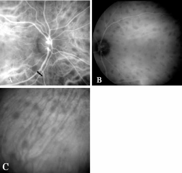
| Fig. 14 A. Mid-phase indocyanine green (ICG) study in a patient with Birdshot choroidopathy. The arrow corresponds to just one of the multiple sites of hypofluorescence caused by the inflammatory lesions in the choroid. B. Late-phase ICG demonstrating multiple hypofluorecent lesions. C. ICG study in the mid-peripheral fundus demonstrating that these hypofluorescent lesions are aligned along the choroidal vessels. |