|
|
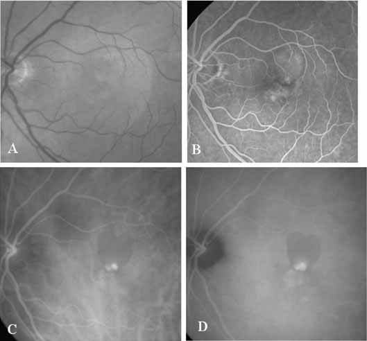
| Fig. 13 A. Clinical photograph of a patient with pigmentary figures and a surrounding retinal pigment epithelial detachment. B. Fluorescein study reveals an indistinct pattern of hyperfluorescence located just below a serous pigment epithelial detachment. C. Indocyanine green (ICG) angiogram demonstrates a hyperfluorescent small polypoidal vessel at the inferior margin of a hypofluorescent retinal pigment epithelial detachment. D. Late-phase ICG study reveals some late hyperfluorescence corresponding to a small plaque inferior to the focal polypoidal lesion seen in C. In this case, the ICG images were used to guide the treatment of the lesion in this patient. Only the hyperfluorescent spots evident on the early-phase ICG were treated. |