|
|
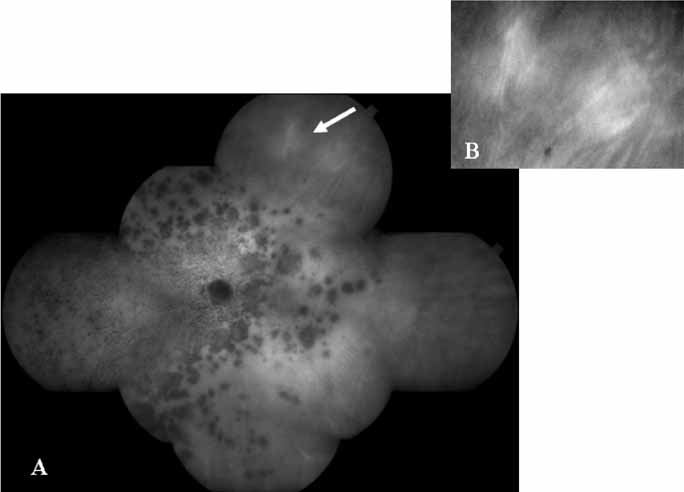
| Fig. 15 A. Composite montage of the ICG angiogram in a patient with multiple evanescent white dot syndrome (MEWDS). Note the numerous large hypofluorescent spots with overlying smaller hypofluorescent dots. These lesions evident on the ICG study far outnumbered the clinically apparent ones. The arrow demonstrates the area that was enlarged in B. B. An interesting finding on the ICG corresponds to these areas of relative hyperfluorescence of the choroidal vessels, which indicate a localized inflammatory reaction. |