|
|
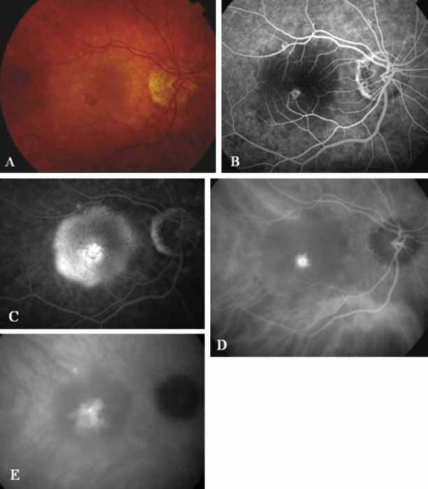
| Fig. 11 A. Clinical photograph of a patient with retinal angiomatous proliferation (RAP). There is a hemorrhage with a retinal pigment epithelial detachment. B. Early-phase fluorescein angiogram demonstrates the retinal–retinal anastomosis. C. Late-phase fluorescein study demonstrates the late-staining hyperfluorescence seen in RAP lesions with a hyperfluourescent retinal pigment epithelial detachment. D. Mid-phase indocyanine green study demonstrates the hyperfluorescent hot-spot overlying the hypofluorescent retinal pigment epithelial detachment. E. Late-phase ICG angiogram demonstrates late hyperfluorescent leakage (CME) into the retina. |