|
|
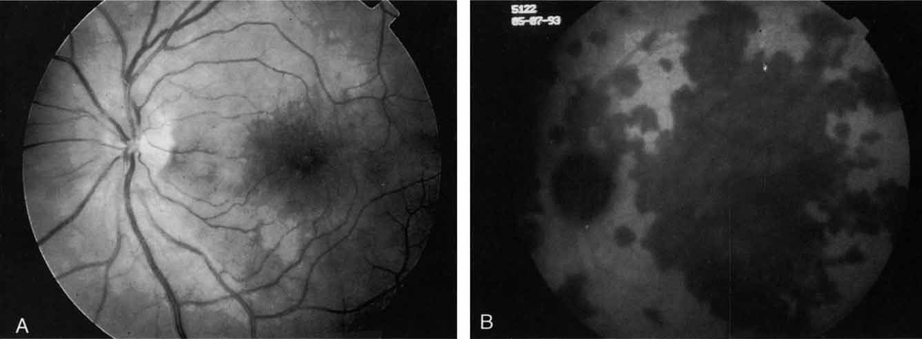
| Fig. 16 A. Clinical photograph of a patient with acute multifocal placoid pigment epitheliopathy (AMPPE). There is extensive involvement of the central macula by the inflammatory lesions. B. Late-phase indocyanine green angiogram demonstrating extensive confluent but irregular areas of marked hypofluorescence. The hypofluorescence in AMPPE is believed to represent a perfusion abnormality within the choroidal circulation. |