|
|
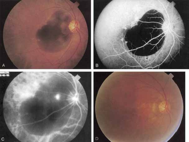
| Fig. 5 A. Clinical photograph of a patient with exudative age-related macular degeneration with secondary subretinal hemorrhage. B. Late-phase fluorescein angiogram demonstrating blocked fluorescence from the subretinal hemorrhage. C. Mid-phase indocyanine green (ICG) study demonstrating blocked fluorescence from the thick areas of subretinal hemorrhage, but a localized area of intense hyperfluorescence representing active choroidal neovascularization was noted in the papillomacular bundle. Laser photocoagulation treatment was applied to this spot only. D. Clinical photograph obtained 3 months later demonstrates an atrophic scar at the site of laser treatment and complete resolution of the subretinal hemorrhage. |