Chapter 19
The Anatomy and Cell Biology of the Retina
SUSANNA S. PARK
Main Menu   Table Of Contents

Search

GENERAL ANATOMY
TOPOGRAPHY OF THE RETINA
RETINAL EMBRYOGENESIS
MORPHOLOGY OF RETINAL NEURONS
GENERAL STRUCTURE OF THE SYNAPSE
NEUROGLIA: OVERVIEW
CIRCULATION
ACKNOWLEDGEMENT
REFERENCES

Interest in retinal anatomy has increased in recent years with the understanding of the importance of retinal function in the visual system. Historically, the retina was first described by Herophilus of Chalcedon (circa 300 B.C.E.) and named as such by Rufos of Ephesus (circa 110 C.E.) because of its appearance as a net to hold the vitreous humor. Despite Galen's early observation of the similarity between this tissue and the brain, little attention was directed to the retina beyond its obvious vasculature and connection to the optic nerve until Kepler (circa 1608) introduced the concept of the retina as the primary photoreceptor tissue of the eye.

With the technical advancement in alcohol tissue fixation, Treviranus first performed the detailed microscopic studies of retinal anatomy in 1835. The later development of thin tissue sections and staining techniques allowed the study of complete neurons and the development of a tentative understanding of the neuronal cell polarity and wiring. The recent use of electron microscopy, immunohistochemistry, and single-cell electrophysiologic studies furthered our understanding of the intraretinal cellular connections and ultrastructure and their role in the complex electrical processing of visual information. New clinical imaging techniques, such as optical coherence tomography, allow us to examine retinal histology in vivo, thus advancing our understanding of pathogenesis of various retinal disorders.

The first detailed study of the anatomy of the retinal vasculature was possible with the advent of the trypsin digest technique. This provided an initial framework for the understanding of retinal vascular physiology. More detailed understanding of the retinal vasculature has been possible recently with progress in whole-mount tissue preparations. Clinical use of fluorescein angiography and newer techniques of vascular flow measurements, e.g., scanning laser Doppler flowmetry, have made possible the correlation between in vivo physiologic vascular changes and previously observed histologic changes in various pathologic entities.

This intimate relationship between the development of concepts of retinal structure and function is no accident. Discoveries in each of these areas have been the basis for further investigation into the other. This chapter is an overview of our current understanding of the retinal anatomy, at both gross and microscopic levels. When appropriate, anatomic correlation is made with various physiologic and pathologic retinal processes.

Back to Top
GENERAL ANATOMY

The retina is a diaphanous, transparent tissue that lines the inner posterior three-fourths of the eyewall.1 It extends from the macula in the posterior pole to a location approximately 5 mm anterior to the equator, the ora serrata, where it becomes contiguous with the nonpigmented epithelium of the pars plana ciliaris. It is loosely adherent to the underlying pigment epithelium, and the two tissue layers can easily be separated in postmortem specimen. The only firm attachments of the retina are at the margins of the optic disc and at the ora serrata (Fig. 1). In living eyes, the retina also is attached to the overlying vitreous in a circular band around the retinal periphery, referred to as the vitreous base. Other points of attachment between the retina and the vitreous may exist around the optic disc and in the macular region.

Fig. 1 Horizontal meridional section of the eye through the central fovea and optic nerve head.

Gross retinal topography reveals characteristic regional variations. The clinical posterior pole is the central posterior retina approximately 5 to 6 mm in diameter, lying between the two temporal retinal arteries (Fig. 2). It is referred to anatomically as the area centralis or macula. Histologically, it is the only region of the retina with more than one layer of ganglion cells. The clinical macula, or the anatomic fovea, is the central area approximately 1.5 mm in diameter within the area centralis. The name macula lutea was given to this region because of the slightly yellow coloration of this area resulting from the presence of xanthophyll in the ganglion and bipolar cells.2 Within the center of the anatomic fovea lies a depression approximately 0.35 mm in diameter surrounded by a ring of slightly thickened tissue. This region is called the foveola by anatomists and the fovea by clinicians. It lies within the capillary-free zone, which measures approximately 0.4 mm in diameter in most people. The photoreceptors in this region characteristically consist of pure cones. The center of the pit within the foveola is called the umbo. It is located 3.4 ± 0.34 mm from the temporal edge of the disc margin and 0.8 mm inferior to the center of the optic disc.3 The anatomic fovea is surrounded by a 0.5-mm-wide ring zone called the parafovea. This area is where the ganglion cell, intranuclear layer, and outer plexiform layer are the thickest. A 1.5-mm ring zone surrounding the parafovea is called the perifoveal area.

Fig. 2 Posterior retina with comparative anatomic and clinical terminologies. (Redrawn from Patz A: Principles of fluorescein angiography. Int Ophthalmol Clin 17:4, 1977)

Beyond the area centralis, the retina spreads out smoothly over the vortex veins, which are visible through the retina and underlying pigment epithelium during ophthalmoscopy. This equatorial region lays approximately 15 mm posterior to the corneoscleral junction and approximately 3 mm posterior to the geometric equator in the emmetropic globe.

At the equator, the retina has a vertical diameter of 24.08 ± 0.94 mm and a horizontal diameter of 24.06 ± 0.60 mm. Measured as a cord, the distance from the corresponding margin of the optic disc to the equator is 14.71 ± 1.08 mm in the superior meridian, 14.51 ± 1.01 mm inferiorly, 13.27 ± 1.11 mm nasally, and 17.29 ± 1.60 mm temporally. This diameter provides a theoretic estimate of the retinal area as 1206 mm2 (estimating the retinal surface to cover two thirds of a sphere of this dimension). Changes in tissue size and shape during preparation have precluded the estimation of the actual retinal area.

Anteriorly, the retina can be seen to terminate in an irregularly wavy edge, the ora serrata. Measured as a cord from the equator, this lies 6 ± 1.22 mm temporally, 5.8 ± 1.12 mm nasally, 5.07 ± 1.11 mm superiorly, and 4.79 ± 1.22 mm inferiorly. Using 1.50 mm as the estimation of a disc diameter (dd), the distance from the equator to the ora is approximately 3 dd. The distance from the anterior limit of the retina to Schwalbe's line is 6.14 ± 0.85 mm superiorly, 6.20 ± 0.76 mm inferiorly, 5.73 ± 0.81 mm nasally, and 6.52 ± 0.75 mm temporally.4 This places the ora approximately at the point of the rectus muscle insertions around the globe. A recent study of the relationship between the ora serrata and the spiral of Tillaux in cadaver eyes showed that in 68% the midpoint insertion of all rectus muscles was within 1 mm of the ora serrata.5 In addition, in most cases, the lateral rectus muscle most consistently inserted closest to the ora.

This anatomic fact is important in vitrectomy surgery. The surgeon can place the sclerostomies for pars plana vitrectomy as far back as 4 mm posterior to the limbus and still have access to the ora serrata extending circumferentially from the meridian of the sclerostomy.6 The 95% tolerance interval for the location of the ora serrata is 4.21 mm posterior to the surgical limbus superiorly and at least 4.81 mm posterior to the limbus temporally.5

The termination at the ora is marked by retinal extensions, labeled teeth, which protrude into the ciliary epithelium. The teeth are more prominent in hyperopic than in myopic eyes, and they have an uneven but characteristic distribution around the ora. Separating the teeth are ora bays, which are indentations of the ciliary epithelium onto the retina. The vitreous base is attached to the retina at the ora from approximately 1.8 mm temporally to 3 mm nasally, posterior to the ora bays.7 The anterior insertion of the vitreous base into the ciliary epithelium is 1 or 2 mm anterior to the ora. The posterior margin of the vitreous base has a relatively smooth contour in normal young eyes, whereas the anterior margin follows the serrations. With aging, the posterior margin of the insertion becomes irregular and migrates posteriorly such that it is close to the equator.8 These changes are thought to play a role in the pathogenesis of retinal breaks from peripheral retinal traction.9

A cross section of a low-power histologic preparation of the retina in the region immediately peripheral to the area centralis shows it to have nine layers (Fig. 3). From internally (the side toward the vitreous), the layers are as follows:

  The internal limiting membrane
  The nerve fiber layer
  The ganglion cell layer
  The inner plexiform layer
  The inner nuclear layer
  The outer plexiform layer
  The outer nuclear layer
  The rod and cone inner and outer segments
  Retinal pigment epithelium

Fig. 3 Light micrograph of human peripheral retina including portion of the choroid (Richardson's methylene blue/azure II stain mixture, 40× objective, Courtesy of Scheie Eye Institute). Inner limiting membrane (arrow): (1) nerve fiber layer, (2) ganglion cell layer, (3) inner plexiform layer, (4) inner nuclear layer, (5) outer plexiform layer, (6) nuclei of photoreceptors (outer nuclear layer), (7) rod and cone inner segments, (8) rod and cone outer segments, (9) pigment epithelium.

Based on histologic studies, the retina is thickest at the foveal margin (0.23 mm) and tapers to approximately 0.10 mm at the foveal center (umbo) and 0.18 mm at the equator. It gradually thins further to 0.11 mm in thickness at the ora serrata (Fig. 4A) as the density of all the neuronal elements, including photoreceptors and ganglion cells, decreases peripherally.3 With use of noninvasive techniques in normal live human subjects, the mean thickness of the foveal center is estimated to be 0.18 mm using the retinal thickness analyzer and 0.15mm with optical coherence tomography (Fig. 4B).10 Among patients with clinically diagnosed macula edema, the mean foveal thickness increases to 0.40 mm using the retinal thickness analyzer and 0.32 mm with optical coherence tomography.

Fig. 4a A. Retina thickness map based on histological studies. The retina is thinnest at the fovea (0.10 mm) and thickest (0.23 mm) in the perifoveal region. Beyond the fovea, the retina rapidly thins up to the equator. Between the equator and the ora, the thickness is relatively constant (0.11 mm). Te, temporal; Na, nasal; OD, right eye. (Straatsma BR, Foos RY, Spencer LM: The retina--Topography and clinical correlations. In New Orleans Academy of Ophthalmology Symposium on Retina and Retinal Surgery. St. Louis: CV Mosby, 1969).

Fig. 4b B. Optical coherence tomography of the macula of the right eye in a healthy 61-year-old man. Macular cross section (i) and thickness map (ii) are in the normal range. NFL (nerve fiber layer); RPE (retinal pigment epithelium).

The distinct layering pattern of the retina reflects the arrangement of neural, glial, and vascular cells that enables the retina to convert and process photic energy into neuronal electrical signals that ultimately reach the occipital visual cortex of the brain. This entire pathway consists of the photoreceptor cell and three neurons.11 The photoreceptor cells (rods and cones) are located in the outermost layer of the retina. The outer segments of the rods and cones initially convert the light signal to electrical energy. The outer nuclear layer contains the cell bodies with nuclei of the photoreceptor cells. In the outer plexiform layer, axons of the photoreceptor cells synapse with the dendrites of the first neuron (the bipolar cell) and with the processes of the integrating neuronal cells, the horizontal cells. The bipolar cells, whose nuclei form the bulk of the inner nuclear layer, synapse in the inner plexiform layer with the dendrites of the second neurons, the ganglion cells. In this layer there are also synapses with amacrine cells, which resemble horizontal cells in that they provide cross wiring within the retinal layer. The ganglion cells extend their axons as the innermost retinal layer, the nerve fiber layer, through the optic nerve to the lateral geniculate body, where they synapse with the third neuron, the cells of the lateral geniculate body, which extend their axons through the optic radiations to the occipital cortex (Fig. 5).

Fig. 5 Neuronal connections in the retina: (1) inner plexiform layer, (2) inner nuclear layer, (3) outer plexiform layer, (4) outer nuclear layer, (5) external limiting membrane, (6) photoreceptor inner and outer segments, (7) pigment epithelium. G, ganglion cells. (Wolff E: The Anatomy of the Eye and Orbit, 4th ed. New York: Blakiston, 1954)

Back to Top
TOPOGRAPHY OF THE RETINA

FOVEA

In the region of the anatomic fovea, the retinal layers have a unique arrangement specialized for color vision and better visual acuity than in the remainder of the retina. The entire fovea measures 1.5 mm in diameter and contains 10% of the cones. It has the highest cone density in the retina, approximately 147,300 per square millimeter.12 The central 0.40-mm zone is free of capillaries and is nourished by the choriocapillaris circulation.13 Clinically, this region is referred to as the foveal avascular zone and can be readily visualized on fluorescein angiography (Fig. 6).

Fig. 6 An arteriovenous-phase fluorescein angiogram shows the narrower lumina of the arterial circulation compared with the venous circulation. Following bifurcation at the disc, both artery and vein form extensive branching networks throughout the retina, except at the avascular fovea. The laminar fluorescein flow evident in the veins is characteristic of the early stage of fluorescein circulation.

In the center of the fovea is a specialized structure 0.33 mm in width, the foveal pit or foveola (Fig. 7).13 This central-most region is free of rods and blue cones (Fig. 8). The red and green cones in this region are oriented parallel to each other and are perfectly straight and vertical with respect to the retinal surface to allow the greatest light sensitivity. The inner segments of these cones are separated from each other by Müller cells.14 In addition, the foveal cones differ from cones elsewhere in the retina. They are long and slender like rods, measuring approximately 80 μm in height. In addition, these cones have a large nucleus and lack the dense collection of mitochondria seen in the ellipsoid of most photoreceptors.

Fig. 7 A. Light micrograph of human anatomic fovea and foveola stained with Richardson's methylene blue/azure II mixture. 10× objective. B. Immunofluorescence labeling of human cones (red with monoclonal antibody against cone-specific enolase) and their nuclei (pink) in the fovea. Note the tightly packed cones with long, thin outer segments and axons forming the fiber layer of Henle (HE). Other cell nuclei in the outer nuclear layer (ONL), inner nuclear layer(INL) and ganglion cell layer (GC) are stained blue with DAPI. Retinal pigment epithelium (RPE) shows auto fluorescence. Foveal pit (*) 10× objective. (Courtesy of Scheie Eye Institute)

Fig. 8 A. Immunofluorescence labeling of foveal cones with anti-red/green cone opsin (red). B. Immunofluorescence labeling of foveal cones with anti-blue cone opsin (red). The majority of cones in the fovea contain red/green opsin. C. Immunofluorescence labeling of fovea with antibody to rod outer segment (red) showing lack of rods in the fovea (*). Other cell nuclei in the outer nuclear layer and inner nuclear layer are stained with DAPI (blue). (Courtesy of Scheie Eye Institute)

To minimize the light scatter through the tissue overlying the cones, the inner central fovea is composed only of processes of the Müller cells. It lacks the inner nuclear layer, inner plexiform layer, ganglion cells, and the nerve fiber layer. In addition, the external plexiform layer has a unique configuration in the anatomic fovea. The external cone fibers (axons) at the foveola make an almost right angle turn away from the foveola and assume a course parallel to the surface of the retina. From the parafoveolar region to the edge of the anatomic fovea, the fibers of the remaining cones and of the rods pursue a similar course. The aggregate of all these horizontally arranged fibers forms Henle's layer of the anatomic fovea or clinical macula (Fig. 7B). After a short course parallel to the retinal surface, these fibers become perpendicular to the retinal surface to synapse with the dendrites of the overlying bipolar cells. This first synapse between the photoreceptors and bipolar cells of the fovea occurs outside the central 0.2-mm diameter of the foveola.14

Clinically, this region has a yellow discoloration on funduscopy because of the presence of xanthophyll in the ganglion and bipolar cells. This macular pigment has several proposed functions, including prevention of chromatic aberration at the fovea, selective absorption of potentially toxic blue light, and quenching of various active oxygen species.15 In the human retina, two components of the macular pigment have been identified.16 They are zeaxanthin and lutein, two structural isomers of dihydroxyxanthophyll. In general, zeaxanthin is confined mostly to the foveal region, whereas lutein is found more widely distributed in the posterior pole. Studies on the monkey retina suggest that a particular ratio of these two xanthophylls may be associated with a specific cone subtype.17 Among patients with age-related macular degeneration, decreased levels of these pigments have been noted in the macula.18 Because these pigments are potent antioxidants, they may play an important role in maintaining the health of the retinal cells in the macula during aging. In animal models, they have been shown to protect photoreceptors from light-induced apoptosis.19

PARAFOVEA

The parafovea, defined as the 0.5-mm-wide annular zone surrounding the fovea (for a total diameter of 2.5 mm), has the largest accumulation of neurons in the entire retina. The bulk of these cells are in the ganglion and inner nuclear layers. This is also the region where the outer plexiform layer forms the thickened Henle's layer by the multitudinous axons of the foveola photoreceptors (see Fig. 7A). This region has a lower density of cones than at the foveolar center. There are approximately 100 cones per 100 μm, and the cone inner segments are separated from each other by 1 to 3 μm.14 The rods also begin to appear in this region, and toward the peripheral edge of this parafoveal zone the outer nuclear layer becomes filled mostly with rod nuclei. Immediately temporal to the papilla, the parafovea contains the highest rod density, 170,000 per square millimeter.12 The inner nuclear layer here is also very dense and consists of up to 12 rows of cells. Similarly, the ganglion cell layer forms up to seven rows of closely packed cells at the foveal edge. The nerve fiber layer is thickest in the region of the papillomacular bundle nasal to the fovea and is thinner temporal to the fovea.

PERIFOVEAL REGION

The perifoveal region is the outermost ring of the anatomic area centralis. It starts at the point where the ganglion cell layer has four rows of nuclei and ends peripherally where the ganglion cell layer is reduced to the single-cell layer, typical of the peripheral retina. Funduscopically, the perifovea includes the circular area 1.25 to 2.75 mm from the foveolar pit. It defines the periphery of the clinical macula, which has a horizontal diameter of 5.5 mm and corresponds to a visual field of 18.3 degrees.

The perifovea differs from the parafovea in that it has a less densely packed array of cone outer segments. The rod density is increased to an average of two rods between adjoining cones. The outer nuclear layer of the perifoveal zone, similar to that of the peripheral parafovea, contains the maximum density of rods. However, the outer plexiform layer and the inner nuclear layer are reduced in thickness in this zone compared with those of the parafoveal zone. This is because the perifoveal outer plexiform layer does not carry the axons of the cones in the foveola. The inner plexiform layer also is reduced to six or seven rows of nuclei as the bipolar cell density decreases away from the parafoveolar zone. In contrast, the inner plexiform layer is slightly thickened, and the ganglion cells are somewhat larger in the perifoveal zone than in the parafovea.

ORA SERRATA

The peripheral edge of the retina is the ora serrata. It marks the junction between the multilayered pars optica retinae and the monolayered nonpigmented epithelium of the ciliary body. The distinct anatomic properties of this region are because of its thinness, lack of vascularity, and intimate relationship to the vitreous base and zonular fibers. The ora serrata receives its name because the retina has marginal notches over much, if not all, of its border with the ciliary epithelium. They are composed of elongations of the retinal tissue into the nonpigmented ciliary epithelium and are known as dentate processes or, more simply, teeth. The excavations between the teeth, which have their convexity posteriorly, are designated as bays. The width of the zone from the tips of the ora teeth to the back of the bays ranges from 2.1 mm on the temporal side of the retina to between 0.7 and 0.8 mm nasally.20

The distribution of ora teeth and bays around the retinal circumference is uneven. The highest density of teeth occurs in the inferonasal quadrant, and the lowest density occurs in the inferotemporal quadrant.3 The number of dentate processes ranges from 16 to 48 among eyes but averages between 25 and 30.20 The processes are longer and thereby more prominent in emmetropic and hyperopic globes than in myopic globes. The teeth are oriented radially with the points aimed between valleys of the ciliary processes. The bays between the teeth may assume various configurations, depending on the depth of the bay and the prominence of the neighboring teeth. The bay may range from only an indentation in the ora to a fully enclosed profile that is surrounded on all sides by retinal tissue. These enclosed bays are anomalies of development and do not change with aging.

The ora teeth may show developmental variations in size and shape.3,21 A regular dentate process is a retinal extension that projects 0.5 to 2.5 mm anterior to the adjacent retina on both sides. In contrast, a large dentate process protrudes onto the pars plana of the ciliary body more than 2.5 mm anterior to the surrounding retina. A giant dentate process extends to or onto the pars plicata of the ciliary body. When a tooth shows thickening by virtue of focal hyperplasia and hypertrophy of its glial component, a solid retinal tuft is evident (Fig. 9). Such a lesion is present in 33% of the adult population. Cystic retinal tufts reflect the same congenital structure as that of the solid tufts, but they are more rare and have a cystic space that results from cavitation within the glial mass (Fig. 10). These lesions have overlying vitreous condensations and may be avulsed by vitreous traction. They occur in approximately 5% of the adult population and may account for 10% of cases of rhegmatogenous retinal detachment. A meridional fold (Fig. 11) is the result of glial overgrowth along a radial line extending anteriorly from behind the base of the tooth to its tip. This is a normal developmental variation that occurs in 26% of the population. A characteristic common to all of these hypertrophic developmental anomalies, except cystic retinal tufts, is that they are benign.

Fig. 9 This noncystic retinal tuft consists of generalized degeneration and gliosis, accompanied by the irregular retinal thinning and pigment epithelial clumping typically found within the vitreous base region. Densely staining glial tissue caps the tuft. (Original magnification ×500; Straatsma BR, Foos RY, Feman SS: Degenerative diseases of the peripheral retina. In Duane TD (ed): Clinical Ophthalmology, Vol 3. Hagerstown, MD: Harper & Row, 1979:1–29)

Fig. 10 Coronal section of an ora serrata dentate process with nonspecific degeneration and microcystic change. The central surface is a cap of dense-staining glial cells that also compose much of the deeper tissue. The ciliary epithelium and the photoreceptors under the dentate process are normal. (Hematoxylin & eosin, original magnification ×250; Straatsma BR, Foos RY, Feman SS: Degenerative diseases of the peripheral retina. In Duane TD (ed): Clinical Ophthalmology, Vol 3. Hagerstown, MD, Harper & Row, 1979:1–29)

Fig. 11 Meridional complexes show the basic constituents: an atypical dentate process that aligns with and extends to an enlarged ciliary process. These complexes also have meridional retinal folds that extend along the dentate process and posteriorly into the peripheral retina. Peripheral retinal excavation (open arrow) is aligned with the complex on left. (Straatsma BR, Foos RY, Feman SS: Degenerative diseases of the peripheral retina. In Duane TD (ed): Clinical Ophthalmology, Vol 3. Hagerstown, MD; Harper & Row, 1979:1–29)

When a dentate process occurs in the same meridian as a ciliary process, a “meridional complex” is formed (see Fig. 11). In this abnormal alignment, the dentate process is exceptionally large and usually combined with a meridional fold. It is continuous with the enlarged ciliary process and often is associated with a peripheral retinal excavation in the corresponding meridian. Both the large dentate process and meridional fold are composed of excessive, disorganized, and somewhat degenerated retinal tissue. Meridional complexes are present in 16% of the population and are bilateral in 58% of affected patients. They are most common in the superonasal quadrant and are multiple in 45% of affected eyes.22

Enclosed and partially enclosed ora bays (Fig. 12) are relatively uncommon developmental variations.23 They are oval islands of pars plana epithelium located immediately posterior to the ora and completely or almost completely enclosed by peripheral retina. These abnormal formations are seen in only 6% of people. In 8% of affected patients, they are bilateral. They are present in 3% of all eyes.22 These formations tend to have the greatest prevalence near the horizontal meridians both nasally and temporally. In 20% of the cases, a meridional fold is found immediately anterior or posterior to an enclosed ora bay.24 The clinical significance of the enclosed bays is that they are often mistaken for a retinal hole. In addition, the posterior edges of enclosed ora bays have an abnormally high rate of retinal tear. After a posterior vitreous detachment, 16% of enclosed or partially enclosed ora bays have an associated retinal tear along the posterior edge.23 This results from the relatively weaker bond of the retina to the underlying pigment epithelium at the edge of the bay as compared with the stronger bond of the retina to the nonpigmented ciliary epithelium. This difference puts excessive traction on the retinal tissue at the posterior margin of the enclosed bay.

Fig. 12 Ora serrata of a 35-year-old man shows an ora bay (island of pars plana) formed by the convergence of two dentate processes into a single one, which has a typical termination between ciliary processes (cp). The posterior edge of the vitreous base is marked by a horizontal line (arrowhead) between the anterior limit of the circulation and the posterior edge of the bays. A posterior peripheral retinal excavation (open arrow) results from localized retinal thinning. (Original magnification ×12; Straatsma BR, Foos RY, Feman SS: Degenerative diseases of the peripheral retina. In Duane TD (ed): Clinical Ophthalmology, Vol 3. Hagerstown, MD: Harper & Row, 1979:1–29)

The ora is approximately 0.11-mm thick and is the thinnest part of the retina outside the fovea (see Fig. 4A). Further thinning within the ora may occur as discrete pits (see Fig. 12). Such pits are frequently aligned with a meridional fold or meridional complex.22 The location of these pits varies from 1 to 7.2 mm posterior to the ora. Their formation is caused by the focal loss of the inner retinal layers (Fig. 13). The tissue surrounding the pit is normal. Such peripheral retina excavations are present in 15% of patients. In 43% of affected persons they are bilateral. Half of the affected eyes contain two or more areas of focal excavation, most commonly in the superior nasal quadrant. Because of an abnormal insertion of vitreous fibers into the retina in meridional folds and complexes associated with pits, the posterior edge of a meridional complex is a common site for a retinal tear. In addition, traction on the retina in the area of a pit could easily produce a full-thickness perforation because of the extreme thinning of the retina at this point.

Fig. 13 A peripheral retinal excavation results from loss of tissue from the inner layers as far external as the inner nuclear layer. The middle retinal layers show degeneration, and the outer layers are intact. (Hematoxylin & eosin, original magnification ×250; Straatsma BR, Foos RY, Feman SS: Degenerative diseases of the peripheral retina. In Duane TD (ed): Clinical Ophthalmology, Vol 3. Hagerstown, MD: Harper & Row, 1979:1–29)

The relationship of the peripheral retina to the vitreous base is important in the development of peripheral retinal tears and rhegmatogenous detachments. The vitreous base normally extends from 2 to 4 mm posterior to the ora (Fig. 14). Within this region, collagen fibrils of the vitreous cortex insert into the internal limiting membrane. With aging, the posterior edge of the vitreous base migrates posteriorly and forms irregular excavations around the globe. The vitreoretinal adhesion is especially strong around this irregular posterior edge.25,26 The significance of this is that the posterior limits of the contour can exert great stress on the retina as the vitreous base retracts forward during aging, leading to a retinal tear at the posterior edge of this adhesion.

Fig. 14 Diagram used for drawing retinal detachments, tumors, and vascular lesions shows the equator as the innermost of the large circles and the ora as the middle circle. Beyond the ora is the pars plana so that the outermost circle represents the posterior edge of the pars plicata. The dimensions in millimeters and in disc diameters refer to the distances from the disc. The fovea is in the geometric center of the diagram. The vitreous base (shaded band) is narrower nasally than temporally. The normal vitreous base extends from posterior to the ora serrata to anterior to all but the longest dentate processes. In senile anterior retraction of the vitreous base, retinal tears may form in the zone between the posterior edge of the vitreous base and the anterior edge of the ora. Te, temporal; Na, nasal; OD, right eye. (Straatsma BR, Foos RY, Spencer LM: The retina—Topography and clinical correlations. In New Orleans Academy of Ophthalmology Symposium on Retina and Retinal Surgery. St. Louis: CV Mosby, 1969)

Histologic Features of the Ora Serrata

The histologic features characteristic of the ora serrata include thinning of the retina caused by a gradual loss of the nerve fiber layer, ganglion cell layer, and plexiform layers (Fig. 15). The outer nuclear layer is reduced to two or three cells in thickness and fused with the plexiform layers.14 This lack of neurons is compensated for by the neuroglia and Müller cells, which serve as the structural matrix for the entire retina. The internal limiting membrane is thickened in a 4-mm-wide zone at the ora because of the insertion of the vitreous base into this region. The rod photoreceptors disappear 1 mm posterior to the ora, where they are replaced by malformed cones. The external limiting membrane continues forward beyond the ora as the layer separating the pigmented from the nonpigmented ciliary epithelium. However, it has morphology different from that of the external limiting membrane of the retina.

Fig. 15 Ora serrata of newborn. The layers develop, in order, from the photoreceptors through the inner nuclear layer to the ganglion cell (open arrow), as the retina recapitulates its embryogenesis in the transition from the nonpigmented ciliary epithelium to the mature retina posterior to the ora. Arrow indicates undeveloped photoreceptors.

A unique feature of the ora is that within its 2-mm width is the transition of the single-layered nonpigmented epithelium of the pars plana into the complex multilayered retina.27 The original light-microscopic observation of the ora suggested that this was a sudden change. Electron-microscopic studies have shown that the transition is gradual and is a recapitulation of retinal embryogenesis (see Fig. 15). In the most peripheral section of the ora, the retinal cells are primitive neuronal precursor cells. These undifferentiated retinal cells at the ora may serve as the storehouse for cells for retinal growth in eyes that grow during early postnatal life or in degenerative myopia. However, the appearance of maturation of the primitive retinal cells at the ora does not always indicate continuous retinal growth, because a similar structure of the ora is seen in eyes of all age groups. Posterior to these precursor cells, 0.28 mm posterior to the ora, undifferentiated photoreceptors are seen. These cells have neither an outer nor an inner segment. In addition, they lack the tight junction connections to the pigmented epithelium that are present between the nonpigmented epithelial cells of the pars plana and the pigmented ciliary epithelium. This explains why retinal detachment usually extends to the border of the ora serrata but only rarely more peripherally.

Farther posteriorly, the photoreceptors take on the configuration of those in the developing retina of a 23- to 36-week-old fetus. The inner segment consists of a mass of cytoplasm protruding past the external limiting membrane. Synaptic connections are occasionally discernible. Slightly more posteriorly, primitive outer segments can be seen. Although they lack the infolding of the plasma membrane characteristic of mature rod outer segments, some of the inner segments in cells have a cilium. Only slightly farther, at the posterior edge of the ora, the photoreceptors assume the adult form found throughout the rest of the retina. The remainder of the neurons make their appearance gradually as the photoreceptors become more mature.

Aging-Related Changes at the Ora Serrata

In an older eye, the histologic changes characteristic of the ora are seen extending more posteriorly than in a young eye. In addition, the ora serrata is subject to degenerative changes with aging. The inner limiting membrane becomes thickened and often vacuolated, and the retinal stroma shows cystoid spaces (Fig. 16). These cysts are usually aligned with dentate processes and are located just posterior to the ora serrata. These cysts may be in the outer plexiform layer (typical cystoid) or in the inner plexiform layer (reticular cystoid). They are formed within Müller cells and neuroglia and are filled with acid mucopolysaccharide. Occasionally, these cysts may communicate with the vitreous cavity or coalesce to develop retinoschisis.

Fig. 16 Retinal termination at the posterior edge of an ora bay shows aging changes of outer plexiform layer cysts (1, 2) and a reticular cyst (asterisk) in the inner plexiform layer, imposed on the characteristic fusion of layers and malformed photoreceptors (arrow). The disappearance of the endothelium and the intramural pericytes of the superficial capillary (open arrow) is characteristic of the obliterated peripheral retinal capillaries, which are marked by their basement membranes.

The physiologic basis for the aging changes in the peripheral retina is unclear. One contributing factor may be the relative avascularity of the ora serrata. The retinal circulation stops approximately 1.5 mm behind the posterior limits of the ora bays, and the ora obtains its nourishment from the choroidal circulation.28 This relative avascularity may result in premature senescence of the retinal tissue of the ora, causing cystoid degeneration to occur.

Back to Top
RETINAL EMBRYOGENESIS

The eyeball starts to develop from neuroectoderm around the twenty-second day of fetal life. A pair of optic primordium, identified as optic pits, form on both sides of the midline in the ventrolateral region of the primitive forebrain. At approximately the 3-mm stage of development, the optic pits extend outward as hollow spheres ventrolaterally from the neural tube to form the optic vesicles.29,30 They become separated from the forebrain by a constriction, or stalk. The cavity of the future third ventricle is continuous with the cavity of the optic stalk and vesicles (Fig. 17A). The next event is the invagination of the optic vesicles, by differential growth and buckling, to form the optic cup. This occurs around the fourth week of fetal life. The optic cup forms a fold inferiorly and ventrally to form the “embryonic fissure” through which the mesenchymal and vascular tissues enter the globe (see Fig. 17A). The embryonic fissure begins to close midway in the fissure and extends anteriorly and posteriorly. The process is completed by the end of the seventh week of gestation. Incomplete closure of the fissure can result in colobomas of the iris, retina, or choroid.31

Fig. 17 A. At 33 days' gestation, embryonic retina is separated from the future pigmented epithelium by the space of the primary optic vesicle (asterisk), which communicates with the cavity of the forebrain (FB) by means of the optic stalks (open arrows). The embryonic or fetal fissure (arrowheads) provides the site of entrance of blood vessels into the optic cup (original magnification ×60). B. At 39 days' gestation, the hyaloid vessels extend from the embryonic fissure through the vitreous cavity to reach the lens. The posterior retina shows differentiation into the neuroepithelium with two distinct neuroblastic layers. The peripheral retina (arrowheads) continues as a monolayer and has not yet formed the bilayered neuroepithelium (original magnification ×150). C. At 35 days' gestation, the retina has the beginning of an inner nuclear zone, the inner neuroblastic layer (1). The outer neuroblastic layer (2) continues to show cell division. The neuroepithelium remains unattached to the underlying multilayered pigmented epithelium. The wide space of the site of the primary optic vesicle (3) is caused by fixation and tissue preparation artifact. The internal limiting membrane is formed and separates the neuroepithelium from the vitreous (V) (original magnification ×300). D. At 41 days' gestation, the neuroepithelium has distinct outer and inner neuroblastic layers. The axons of the ganglion cells have begun to form the nerve fiber layer as they migrate toward the embryonic fissure, where they will form the optic nerve (arrow). V, vitreous body (original magnification ×200). E. At 10 weeks' gestation (54 mm), the division into the two neuroblastic layers has extended peripherally up to the future ora serrata (arrowhead). Retinal maturation progresses from the posterior pole to the anterior region of the optic cup and from the inner ganglion cell layer outwardly to the photoreceptors. L, lens; V, vitreous body (original magnification ×100).

The retina develops from the neuroectoderm following the formation of the optic cup. The inner layer of the optic cup develops into the retina, and the outer layer develops into the retinal pigment epithelium. The differentiation of multipotent stem cells of the single-layer optic vesicle ectoderm into the multicellular mature retina is an ordered event that follows discrete stages. Recent studies indicate that cell fate determination in the vertebrate retina depend on changes in the response of the mitotic cells within the retina as well as changes in environment around these cells.32 The first stage is the differentiation of the cells of the optic vesicle into a two-layered tissue, the “neuroepithelium.” This stage of development commences simultaneously with the formation of the optic cup. During this process, a layer of ependymal cells that show numerous mitoses appears at the edge of the vesicle. At the base of these cells is a basement membrane from which extend numerous fine cilia.29 Interdigitation of the cilia with the cells of the outer layer of the optic vesicle is a precursor to the subsequent arrangement of the insertion of the photoreceptor outer segments into the spaces between the microvilli of the pigmented epithelium. Immediately internal to the ependymal layer is the thick layer of primitive undifferentiated cells, which are characterized by cytologic evidence of high mitotic and metabolic activity—large oval nuclei with darkly staining nucleoli and a prominent chromatin net. The ependymal layer and the aggregation of the nuclei of the undifferentiated cells form the “primitive zone,” constituting the outer 90% of the neuroepithelial thickness. The inner 10% is made up of the “marginal zone,” which consists of the cytoplasmic extensions of the multitudinous outer primitive cells. The marginal layer is microscopically differentiated by its lack of nuclei until the 10-mm stage at 6 weeks of development.

At the beginning of the invagination of the optic vesicle, the distal end of the primitive ophthalmic artery grows into the embryonic fissure from below. When the fissure closes, the artery becomes trapped within the cavity separating the marginal zone from the lens vesicle. Intraocular branches of this transitory hyaloid artery (Fig. 17B) presumably meet the nutrient requirements of the developing retina until its own vasculature is established during the fourth month of gestation.

From the sixth week to the third month, the neuroepithelium gradually develops into the neuroblastic layers, from which the mature retina subsequently develops. The pattern of this change resembles the morphogenesis of the central nervous system, with the spread of cellular proliferation directed from the inner layer toward the outer layers. Because of its invagination, the neuroepithelium's direction of proliferation is translated into progression from the outer toward the inner layers (Fig. 17C). The primitive cells internal to the ependymal layer divide and migrate inward to form a distinct new layer of nuclei where the marginal layer had been (see Figs. 17B and 17C). This results in the formation of the inner and outer neuroblastic layers, which are separated by the transient nerve fiber layer of Chievitz (see Figs. 17B and 17D). This distinct, three-layered structure is temporary. It gradually takes on the more complex architecture of the retina by cellular differentiation. This differentiation of the neuroblastic layers progresses from the inner layer (future ganglion layer) to the outer layer (future photoreceptor layer) such that the outer retina takes on its mature appearance last.33 In addition, this differentiation of the neuroblastic layers is more precocious in the posterior pole as compared with the periphery (see Figs. 17B and 17E), and the development of the supportive structures occurs earlier than that of the neural structures.

Exemplifying these developmental principles is the finding that the Müller cells, which are the primary supportive elements of the retina, are the first of the retinal cells to appear, and they are found at the posterior pole as early as the 10-mm stage. The original position of the Müller cells is adjacent to the internal limiting membrane, which they contact with their inner processes. This results in a double-layered internal limiting membrane whose inner component is the basement membrane formed by the original neuroectodermal cells, and the outer layer is the pseudomembrane formed by the adhesion of the terminal processes of the Müller cells. The Müller cells elongate to extend through the full thickness of the retina until they insert onto the outer basement membrane, which was previously formed by the neuroectodermal cells. The external limiting membrane forms when the Müller cells develop junctions with their contiguous neighbors. Emerging evidence indicates that the molecular basis for regulating Muller cell development may depend on intracellular and extracellular signaling pathways. The Notch, a contact-mediated signaling pathway, as well as cyclin dependent kinase inhibitors have been implicated to play a role.34

Once Müller cell differentiation occurs, the third stage of retinal development follows with the differentiation of the neural elements. The inner neuroblastic layer undergoes this differentiating process before the outer layer does (Fig. 18A and 18B, top). The ganglion cells migrate inward from the inner neuroblastic layer, creating a gap between them and the remainder of the inner plexiform layer. The nerve fiber layer becomes the first mature retinal layer;35 it gradually forms from the 17-mm (see Fig. 17D) to the 130-mm stage as the ganglion cells send their axons toward the optic disc.

Fig. 18 A. In the 24-mm human embryo (approximately 7 weeks' gestation), the developing retina is divided into two layers. The nuclei of the cells of the inner neuroblastic layer (1) are oriented in various directions without any layering. The layer of Chievitz (2) is composed of interconnecting processes and also contains nuclei that are migrating from the outer toward the inner retina. The closely packed cells of the outer neuroblastic layer (3) have nuclei of variable densities. Mitoses (arrowhead) occur in the outer layer. The underlying pigmented epithelium (PE) cells show clumping of their granules at their apical ends (original magnification ×720). B. In the retina of the 50-mm fetus (10 weeks' gestation), cells of the inner neuroblastic layer (1) have started to differentiate. Immature ganglion cells (arrowheads) are near the inner limiting membrane. The layer of Chievitz (2) contains some cells presumably in transit between the two nuclear layers. (3) Outer neuroblastic layer. The future cone nuclei (arrows) are positioned near the monolayered pigmented epithelium (original magnification ×720).

The next step in differentiation occurs between the 17-mm stage and the fourth and fifth months of development. The inner nuclear layer is created by inward migration of a part of the outer neuroblastic layer and joining of these cells with the amacrine and müllerian cells of the inner neuroblastic layer (see Fig. 18A and 18B, bottom). This results in the obliteration of the layer of Chievitz and the formation of the outer plexiform layer, which occur during the 10th and 12th weeks of gestation. These developmental changes occur from the posterior pole to the periphery of the retina, with the exception of the macula, which is the last region to undergo development.36 The remainder of the outer neuroblastic layer cells, which are precursors to photoreceptor cells, become a row of cells with the nuclei internal to the outer limiting membrane by the fifth to sixth month. This establishes the retinal layers that will persist into maturity. However, the morphologic development into primitive photoreceptor cells does not begin until the 48-mm stage (see Fig. 18A and 18B, bottom).

The photoreceptor cell bodies derive from these late-developing outer neuroblastic cells, and the photoreceptor outer segments arise from cytoplasmic processes that either originate from or replace the cilia of the original outer membrane at the 17-mm stage.29 These processes rapidly form interdigitations with the microvilli of the pigmented epithelial cells. At a far later time, at approximately the 80-mm stage, cells of the outer nuclear layer extend protoplasmic processes that penetrate the original external basement membrane and form the inner portion of the photoreceptor outer segments. Thus, the photoreceptor outer segments form developmentally as a separate unit and become connected to the remainder of the photoreceptor cell by a narrow stem later in embryogenesis.

The genesis of retinal architecture follows a spatiotemporal gradient probably regulated by local interactions.37,38 Action potentials endogenously generated by the ganglion cells, the first cells to differentiate in the neural retina, appear to be necessary for development of the retinal layers.39 Spontaneously generated calcium waves have been detected in ganglion and amacrine cells of the immature retina before photoreceptor development. These waves are synchronized and spread horizontally in a wavelike manner across the retina.40 They are hypothesized to refine the neural connections and confer topographical mapping to retinal connections. The temporal characteristics of these calcium waves appear to be regulated by GABA, a neurotransmitter expressed by human fetal retina as early as 12 weeks of gestation.41,42

The formation of the vascular system in the internal retina by the eighth month marks the end of retinal maturation, except for foveal development, which continues until 6 months after birth. Any growth of the retinal area beyond its size at the eighth month of fetal development results from thinning of the existing structure, because there is no further mitotic proliferation or cellular hypertrophy of retinal neurons after that time. This accounts for the peripheral retinal thinning characteristic of myopic eyes in which globe enlargement occurs years after completion of the tissue's growth potential. Similarly, the normal retina cannot regenerate after destruction of retinal tissue by infection, inflammation, vascular insufficiency, surgical photocoagulation, diathermy, or cryotherapy. In these cases, retinal replacement occurs only through glial cell metaplasia and hyperplasia.

The macula is the last retinal region to reach morphologic maturity.29,30 The macula retains the transient layer of Chievitz until 4 months after birth. The ganglion cells continue to accumulate in the macula until the sixth month of fetal life, with no evidence of the tissue thinning that characterizes the growth of all other retinal regions. Studies in monkeys indicate that the formation of the foveola and the development of the morphology of cells within the foveola depend on the formation of high densities of retinal ganglion cells within the central retina.43 The foveal pit results from the gradual loss of overlying ganglion cells as they spread slightly peripherally during months 7 and 8. Similarly, the amacrine, horizontal, Müller, and bipolar cell nuclei overlying the center of the fovea are displaced.44 Until 4 months after birth, the macula continues to mature with elongation and full development of the cones and peripheral migration of the remaining ganglion and bipolar cells. The result is the disappearance of the layer of Chievitz and the formation of the foveal pit. This late maturation of the fovea accounts for the delay in development of fixational ability until the postnatal age of 4 months.

Back to Top
MORPHOLOGY OF RETINAL NEURONS
The cells within the retina can be divided into neuronal, glial, and vascular components. The neuronal cells are responsible for the primary function of the retina (i.e., conversion and transmission of incoming light into an electrical signal perceived by the brain). This is achieved through intricate synaptic connections among three different classes of retinal neurons, namely, photoreceptors, interneurons, and ganglion cells. A detailed understanding of the cellular morphology of these neuronal cells and their synaptic connections has been possible in recent years with the advent of electron microscopy, immunohistochemistry, and Golgi-impregnated preparations. Morphologically, 55 different types of retinal neurons have been identified in the mammalian retina to date. The function of 22 of these cell types is known. Each morphological type of retinal neuron appears to have a distinct physiological function. As further studies improve our understanding of the retina's role in processing the visual signal, the function of the remaining identified cell types will become apparent.
Back to Top
GENERAL STRUCTURE OF THE SYNAPSE
To appreciate the complexity of the interneuronal connections within the retina, an understanding of the basic morphology and function of synapses and other interneuronal junctions is important. The retinal neurons form three major types of intercellular connections: the chemical synapse for neuronal signal transmission, the zonula adherens that maintains tissue structure, and the gap junction that provides electrical transmission between developing retinal cells. These junctions have important morphologic similarities and differences that reflect their differences in function.

The synaptic junction of a typical chemical synapse consists of specialized apposed membranes of the two neuronal cells, separated by a 20- to 30-mm cleft.45 Similar to other intercellular junctions, such as zonulae adherentes, the plasma membrane of the synaptic junction contains tufts of fine filaments that insert into cytoplasmic densities, and the two apposing cell membranes are glued together by a thin, dark plate of filamentous material in the intercellular cleft.46 However, unlike the interepithelial cell adherence, the interneuronal synapse is polarized and has an asymmetric predominance of intracytoplasmic fibrils in the postsynaptic side of the junction.

The distinction between the morphology of the synaptic junction and that of the zonula adherens is important because both types of junctions are present between neuronal cells in the retina. These nonsynaptic junctions do not serve the purpose of electrical communication between cells. As in epithelial tissue, where they are commonly found, the zonulae adherentes act as binding sites to maintain tissue cohesion.45 In neural tissue they are usually short and have been given the special label puncta adhaerentia.46

Another type of junction between neuronal cells in the retina is the gap junction.47 In a gap junction, there is close apposition between presynaptic and postsynaptic membranes, and current is allowed to pass directly between adjacent cells. Thus, it is classified as an electrical synapse. Because no chemical mediator substance is involved, the rate of signal transmission is faster in these junctions than in typical chemical synapses. In some synapses, characteristics of both the chemical synapse and the gap junction are found. These connections, called “mixed synapses,” have not been found in the retina thus far.45

The electrical synapse (gap junction) characteristically shows a transsynaptic gap with a seven-layered structure produced by the approximation of membranes from the two juxtaposed cells (Fig. 19).48 The synapse consists of four dense lines and three intervening light spaces. It lacks the morphologic detail characteristic of the chemical synapse. Thus, they are localized best by tracer coupling. There are no vesicles and no obvious changes in the cytoplasm on the two sides of the junction. The gap junction does not function by the use of a chemical transmitter. Instead, it offers a low-resistance pathway between the cells. The extent of the electrical coupling of the adjacent cells appears to be dependent on the level of expression and type of the connecting particles (connexin). Thus, plasticity of the coupling in the retina is possible and the retina has the potential to optimize vision for the ambient lighting conditions.49

Fig. 19 Electron micrograph of a gap junction between two horizontal cells in a teleost retina. (Courtesy of Dr. Charles Zucker)

The close apposition of the plasma membranes of the gap junction allows for rapid and well-insulated electrical transmission to occur in either direction. Gap junctions have been identified between photoreceptor terminals, horizontal cells, and amacrine cells.50–54 In the vertebrate retina, gap junctions usually mediate lateral connections, coupling neurons of a single subtype into an extended array of mosaics in the plane of the retina (homologous).55 Occasionally, these electrical synapses can be seen between bipolar terminals and amacrine cells processes (heterologous).56,57 In these instances, they may play a role in transmitting signals from the photoreceptor to the ganglion cell.58 Based on biotinylated tracer studies, at least 3 different types of gap junctions have been found.59 The most permeable gap junctions were between A-type horizontal cells. The lowest permeability gap junctions were found between cone bipolar cells and AII amacrine cells. Intermediate range gap junctions were found between amacrine cells and between B-type horizontal cells.

In the retina, the major type of interneuronal junction of interest is the chemical synapse. In such a synapse, intercellular communication is achieved when a chemical substance, the neurotransmitter, synthesized from the presynaptic axon terminal, is released into the synaptic cleft, where it binds to specific receptors on the postsynaptic membrane. This binding of the neurotrans-mitter elicits an electrophysiologic change in the postsynaptic neuron that may be excitatory or inhibitory.60 This response, in turn, can be affected by neuromodulators released by neurons that can alter the metabolism of the neurotransmitter or the postsynaptic electrophysiologic response. Table 1 summarizes the putative neurotransmitters and neuromodulators that have been associated with presynaptic terminals of various retinal neurons.60–62 Immunohistochemical studies show that each of these neurotransmitters may be associated with only a subpopulation of a type of retinal neuron.63 In addition, there may be co-localization of more than one neurotransmitter in the same retinal neuron.62,64 In photoreceptors, only glutamate and aspartate have been identified as neurotransmitters. Colocalization of four or more neurotransmitters is often observed in the interneurons.64,65


TABLE 1. Putative Neurotransmitters and Neuromodulators of the Retinal Neurons


Presynaptic NeuronNeurotransmitter/NeuromodulatorEffect on Postsynaptic Neuron
PhotoreceptorsL-Aspartate/glutamateExcitatory—type I horizontal cell
 TaurineNeuromodulation—photoreceptor
 Melatonin 
Horizontal cells (type I)GABA/glycineInhibitory
 L-Aspartate/glutamateExcitatory
Bipolar cellsL-Aspartate/glutamateExcitatory
Amacrine cellsGABAInhibitory
(Starburst)GlycineInhibitory
 DopamineExcitatory or neuromodulation
 L-Glutamate/aspartateExcitatory
 TaurineInhibitory
 SerotoninN/A
 EpinephrineExcitatory
 AcetylcholineExcitatory
 NeuropeptidesExcitatory/Inhibitory
 Enkephalin 
 VIP 
 Substance P 
 Glucagon 
 Somatostatin 
 LH-RH 
Interplexiform cellsDopamineExcitatory/Inhibitory
 GlycineInhibitory
 GABAInhibitory
Ganglion cells >L-Glutamate Excitatory
 L-AspartateExcitatory
 Acetylcholine 

GABA, γ-aminobutyric acid; VIP, vasoactive intestinal polypeptide, LH-RH, luteinizing hormone-releasing hormone.
Data from multiple souces.60–62,64,65


The basic feature of the presynaptic elements of the chemical synapse is the boutons terminaux at the ends of the axons, or boutons en passant along their length.45 In the central nervous system, these occur at points where myelination has ceased. The lack of myelination in the retina makes it impossible to use myelination gaps for synapse identification. Although most synapses are axodendritic, axosomatic, or axoaxonal, other types have been documented in the brain and the retina where the presynaptic component is not axonal but dendritic or somatic. However, all of these synaptic types share a common morphology consisting of presynaptic and postsynaptic membranes, the densities associated with their cytoplasmic faces, and the synaptic cleft between them.

In the retina, three different classes of chemical synapses have been described based on the appearance of the presynaptic element: conventional synapse, ribbon synapse, and flat or basal junction.66 The conventional synapse, which is similar in morphology to other chemical synapses found throughout the vertebrate nervous system, has aggregates of synaptic vesicles clustered close to the presynaptic membrane. These vesicles are membrane-bound organelles that contain neurotransmitter substances and enzymes involved in neurotransmitter metabolism.67 They lie in the midst of a hexagonal array of electron-dense particles, each 30- to 50-nm wide and separated from neighboring particles by an interval of 20 to 50 nm.68 This electron-dense material is believed to play a role in the attachment of the synaptic vesicles to the presynaptic membrane. These synapses are found in the presynaptic terminals of horizontal cells, amacrine cells, and interplexiform cells.66 They typically have an inhibitory effect in the postsynaptic neuron.

The ribbon or invaginating synapse, in contrast, is usually found in the presynaptic terminals of photoreceptors and bipolar cells in the retina (Fig. 20). They are also often observed throughout the invertebrate nervous system. The presynaptic terminal of the ribbon synapse is characterized by the presence of an electron-dense bar or a ribbon of approximately 1 um length that is oriented perpendicular to the presynaptic membrane. Reconstruction reveals that this ribbon is in fact a rectangular or horseshoe shaped plate composed of two osmiophilic electron-dense lamellae flanking an electron-dense central layer69,70 This synaptic ribbon is surrounded by a precisely arranged array of synaptic vesicles and appears to be anchored to the presynaptic membrane by three to five delicate electron-dense filaments. The shape and size of the synaptic ribbons can change depending on the maturation stage, activity state of the synapse, or light/dark cycle. Although the precise function of the synaptic ribbon is unknown, it has been hypothesized to function as a “conveyor belt” to channel synaptic vesicles to the presynaptic membrane for neurotransmitter release, to immobilize the vesicles in inactive synapses, storage device for synaptic transmitter, and to act as a diffusion barrier for presynaptic ions.69 In the inner plexiform layer, this synapse has two postsynaptic processes, forming a “dyad.” In the outer plexiform layer, there are three postsynaptic processes, forming a “triad” (see Fig. 20).

Fig. 20 Two ribbon synapses in a rod spherule (S) are surrounded by synaptic vesicles: (1) central bipolar dendrite terminal and (2) terminal buds of horizontal cells. Arrowhead points to arciform density (original magnification ×24,000).

Basal junctions are found in the presynaptic terminal of photoreceptors only.66 In the mammalian retina, these junctions are found only in association with cone cells. Typically, the presynaptic membrane is smoothly indented, and there is prominent electron-dense material on the inner surface of the terminal membrane. However, no synaptic vesicle is seen in association with the electron-dense material. The exact mechanism for chemical transmission through these synapses is unclear. However, in axonal terminals of goldfish horizontal cells in which no presynaptic vesicles are found, neurotransmitter release is believed to occur by a carrier-mediated mechanism.71 Such a process may be involved in basal junctions of mammalian photoreceptors. The involvement of a neurotransmitter in signal transduction is implicated by freeze-fracture studies that show that the postsynaptic membranes of these synapses contain particles that are similar to those found in the postsynaptic membranes of these synapses contain particles that are similar to those found in the postsynaptic membranes of a conventional synapse in the brain.

The synaptic cleft of a typical chemical synapse is 20- to 30-nm wide.45 It is not a free space because it houses material that binds the presynaptic and postsynaptic membranes together in a strong adhesion. The electron-dense plaque in the middle of the synaptic space contains mucopolysaccharides and proteins, which may serve to maintain the attachment between the presynaptic and postsynaptic membranes through a form of polyionic binding. Filaments composed of macromolecules span the synaptic cleft. This synaptic cleft substance may be important for formation, localization, and proper function of the synapse. Macromolecules spanning the gap between the presynaptic and postsynaptic membranes may ensure rapid transport of the transmitter molecule through the cleft and may prevent the transmitter from diffusing away from the synapse.72

In the postsynaptic membrane, a linear density is present. From this structure, filaments project out toward the cytoplasm.45 These densities form either a single large or several small discs, which may be interspersed with puncta adhaerentia. The anatomic location of the receptors for the transmitter molecule in the postsynaptic membrane has not yet been determined. However, freeze-fracture studies have shown minute bumps within the specialized zone of the postsynaptic membrane that may represent the neurotransmitter receptor molecule.66 In the case of the neuromuscular junction, alpha-bungarotoxins that bind to the acetylcholine receptor have been localized in the regions of the postsynaptic membrane. It presently is believed that the specialized region of the postsynaptic membrane contains the receptor-ionophore complex responsible for the postsynaptic changes in ionic permeability.73 Consistent with this hypothesis, electrophysiologic studies have shown that there is a sharp, focal increase in sensitivity to the transmitter in the postsynaptic membrane bouton, suggesting that the receptors for the transmitter are present in high density in this region.74 However, this postsynaptic specialization may be reversible, because this localized distribution of transmitter receptor activity is lost and receptor activity is detected in the extrasynaptic membranes of the postsynaptic cells when presynaptic neuronal input is inhibited.

Extensive research has been done recently to identify receptors for the neurotransmitters in the postsynaptic neurons of the retina. The neurotransmitter receptors are extremely diverse in their subunit composition and their exact actions and the signaling pathways on the postsynaptic neurons are not fully elucidated. Glutamate receptors have been identified on bipolar, horizontal and amacrine cells.75 Glycine receptors have been identified on ganglion and bipolar cells.76–78 GABA receptors have been found on photoreceptors, bipolar cells and ganglion cells.78–80 Nicotinic acetylcholine receptors have been found on amacrine and ganglion cells.81 Purinergic receptors have been found on bipolar cells.82 Dopamine and somatostatin receptors are found on all retinal neurons.65,83 However, most of the dopaminergic actions appear to be extrasynaptic. Further studies are needed to complete our understanding of the role of these neurotransmitters in the visual signal processing of the retina.

PHOTORECEPTOR CELLS

The photoreceptor cells of the retina consist of the rods and cones. These are the primary neurons in the visual pathway. Because they derive embryonically from the ciliated cells lining the optic vesicle cavity, they come to lie at the outer edge of the retina. The dense packing of the photoreceptors, as well as their precisely axial arrangement relative to the optical system of the ocular media, provides for detailed sampling of the visual field. Any change in the arrangement of these long, narrow cells so that they are no longer axial with the impinging light results in alteration in vision. Micropsia results if the photoreceptors are abnormally separated from each other but maintain their normal alignment axial to the optical system. This can occur with central serous retinopathy. Metamorphopsia results if the alignment is partially lacking, as in cellophane maculopathy or serous elevation of the macula. Loss of visual acuity can result if the alignment is sufficiently disturbed so that the photoreceptors are no longer relatively axial to the impinging light and lose their sensitivity.

The photoreceptor cells differentiate longitudinally into four major regions: the inner segment containing the metabolic apparatus, the outer segment containing the visual pigment for the conversion of light into neuroelectrical energy, a perikaryal region containing the cell nucleus, and a synaptic terminal.11 The inner and outer segments of both rods and cones are connected by a thin cytoplasmic bridge containing a cilium (Fig. 21).

Fig. 21 Electron micrograph of portions of rod inner and outer segments: connecting cilium (cc) between rod outer segment (os) and ellipsoid (El); basal body (centriole; open arrow); striated ciliary rootlets (arrowhead) between long mitochondria of inner segment; mucopolysaccharide–protein-containing matrix (im). Note rod saccules (discs) covered by plasma membrane (solid arrow). Asterisk indicates basal rod discs in formation (original magnification ×24,000). Inset. Schematic drawing of retinal receptors and interreceptor contacts. Rod spherule is in the middle. (Sjöstrand FS: Electronmicroscopy of the retina. In Smelser GK (ed): The Structure of the Eye. New York: Academic Press, 1961:1–28)

Histologically, the photoreceptors are classified into two types: rods and cones. They differ in the shape of the inner and outer segments, position of the nucleus, and shape of the synaptic terminals (see Fig. 21). The outer segments in rods and foveal cones are cylindrical, whereas those in extrafoveal cones are tapered.11 In general, a cell with the cone-like outer segment is associated with a large synaptic terminal, called a pedicle. The synaptic terminals of rods are smaller and are called spherules (see Fig. 20).

In addition, rods and cones have a different topographic distribution in the retina. Although the rods far outnumber the cones in the total retina, with a ratio of 125 million rods to 6.5 million cones, the cones are present predominantly in the foveal region, where they reach a density of 150,000 per square millimeter.12 The cone density decreases to 75,000 per square millimeter at 130 μm from the foveal center, at which point the rods begin to appear. Approximately 3 mm from the foveal center, the rods are present at their highest density, whereas the density of the cones continues to diminish.

Retinal photoreceptor density decreases with increasing age in humans. The annual cell loss is approximately 0.2% to 0.4%, similar to age-related loss of retinal ganglion cells and retinal pigment epithelial cells.84 Rods are more affected than cones. The decline in density is more pronounced 5 to 8 mm from the foveal center than the retinal periphery. All quadrants of the retina appear to be equally affected. This decline may play a role in age-related retinal disorders. In patients with age-related macular degeneration, an even more pronounced loss of the rods are noted in the parafovea when compared to age-related controls.85 Recent immunohistochemical studies of postmortem eyes indicate increased apoptosis in the macular photoreceptors in patients with atrophic macular degeneration (Fig. 22).86 In patients with advanced macular degeneration, virtually all surviving photoreceptors in the macula are cones, a reverse of the normal macula which contains mostly rods.85 Similarly, among patients with retinitis pigmentosa, mutations in proteins of the photoreceptor outer segments appear to selectively cause apoptosis of rods. Cones do not appear to be directly affected by the mutation but appear to die secondary to rod cell death. The mechanism of cone cell death in retinal degeneration is unknown at present.

Fig. 22 Immunofluorescence photomicrograph of a retina section from a post mortem donor with geographic atrophy from age-related macular degeneration (AMD). Nuclei are labeled with DAPI (blue). Retinal pigment epithelium (RPE) is yellow as a result of autofluorescence. At the edge of atrophy, RPE disappears first and photoreceptors form a psuedorosette. Photoreceptors label with anti_Fas receptor (red, asterisk), suggesting that the Fas pathway may participate in photoreceptor death in AMD. Dying photoreceptors whose nuclei are TUNEL positive (green-arrow) are present consistent with apoptosis. Outer nuclear layer (ONL), inner nuclear layer (INL), ganglion cell layer (GCL). Scale bar 50 microns. (Courtesy of Joshua L. Dunaief, MD PhD, Scheie Eye Institute, University of Pennsylvania)

Outer Segments

The outer segments of rods and cones differ in their size, shape, and structure. Rod outer segments are 25- to 28-μm-long cylindrical structures with a diameter of 1 to 1.5 μm. The outer segments of extrafoveal cones are shorter and tapered—the diameter decreases from 6 μm at the base to 1.5 μm at the tip. The outer segments of foveal cones are tightly packed and elongated and appear more like rod outer segments.87,88

Both rod and cone outer segments contain an elaborate system of stacked membranous discs that arise from invaginations of the cell membrane during development. The main morphologic difference between the rod and cone outer segments is that these discs are continuous with the cellular membrane of the outer segments in cones, whereas in rods they have no continuity with the surrounding cell membrane except at the base of the outer segment.89 Despite this “free-floating” appearance of the rod outer segment discs, they are held in place by a filamentous cytoskeletal structure that spans the rims of these discs.90

The membrane of the outer segment discs contains 90% of the photopigment.11 Each outer segment disc of rods contains 300 to 900 molecules of the photopigment.90 In rods, the visual pigment called rhodopsin is composed of the light-absorbing chromophore retinal, which is attached to a protein moiety called opsin.91 This photopigment is most sensitive to light that is approximately 500 nm in wavelength. Similar photopigment has been found in cones. However, there are three different types of cones, and each cone type contains a different protein moiety in the photopigment. This results in three different spectral sensitivities among the cone photopigments. The blue-sensitive cone pigment has maximal absorption of light at approximately a 450-nm wavelength, the green-sensitive cone absorbs at approximately 530 nm, and the yellow-sensitive cone absorbs at approximately 565 nm. Defects in color vision result from absence or dysfunction of one or more of these cone types.

During the 1960s, Young showed evidence that the rod outer segments are constantly renewed.92 Subsequently, it has been shown that cones also shed their discs.93 In rods, the sacs are formed in the inner segment of the photoreceptor cell and shed at the outer segment tip. These shed sacs, in turn, are phagocytosed by the adjacent pigment epithelial cell (Fig. 23). This cycle of disc shedding has been found to be a circadian phenomenon in some species. In others, it appears to be triggered by light. The physiologic factors that regulate the cycle of disc shedding are still unknown and under investigation.

Fig. 23 Main figure. The rod outer segment (R) with discs is surrounded by pigment epithelial villi (arrowheads) and pigment granules (Pi) from the enveloping pigment epithelial cell (original magnification ×48,000). Inset. The interdigitation of the pigment epithelial cells (I) and the rod outer segments (2) places the outer segments near the apical pigment granules. (3) Inner segments. The external limiting membrane (arrowhead) lies internal to this region (original magnification ×720).

The formation of photoreceptor discs in rods begins with the synthesis of rhodopsin in the rough endoplasmic reticulum and Golgi apparatus of the inner segment.94 Here, the protein is inserted into the lipid bilayer of the endoplasmic reticulum, which eventually fuses with the apical inner segment plasma membrane. The protein is thought to diffuse in the plane of the cell membrane and become incorporated into new disc membranes formed at the base of the outer segment.

When the sacs first appear in the base of the outer segments, they are immature, with poorly oriented discs and membranous tubules. As these sacs increase in number and size, they move distally and take on a mature appearance as they become oriented parallel to the older sacs. The entire progression down the outer segment requires approximately 9 to 13 days. New discs are formed at the rate of approximately 90 per day.95 When the sacs reach the distal end of the rod outer segment, their tips detach and undergo phagocytosis by the adjacent pigment epithelial cell. Recent evidence indicates that a disturbance of the pigment epithelial cell's phagocytic capacity may be a factor in the retinitis pigmentosa class of retinal degenerations.93 Whether this is caused by a primary malfunction of the pigment epithelial cell or to an abnormality of the outer segment that prevents normal phagocytosis is unknown. In studies of dystrophic rat retinas where there is a defect in phagocytosis, the primary defect in gene expression appears to be at the level of the retinal pigment epithelium.96

The discs of cones have a pattern of growth different from that of rods. Radioactive precursor studies show that new protein is incorporated into the cell membrane throughout the outer segment, rather than just at the base.97,98 This implies that cone outer segment renewal is a constant process that is under way at many points throughout the outer segment. This correlates with the previously mentioned structural difference between the rods and cones, wherein cone discs are actually invaginations of the cell membrane, and the rod discs appear to be completely separate from the cell membrane except at the base.

The photoreceptor outer segments have two important connections. One of these is to the inner segment, which is actually the cell body of the photoreceptor (see Fig. 21). The other connection is to the extracellular matrix that separates the photoreceptor outer segment from the pigment epithelial cell. Radioactive tracer experiments suggest that the sources of this extracellular matrix substance are the pigment epithelial and photoreceptor cells.99,100 The material, a glycoprotein consisting of chondroitin sulfate and sialic acid, is secreted into the space between the outer segments and the pigment epithelial cells by vesicles that originate in both cells. The function of this mucopolysaccharide-rich matrix is not well understood. It may provide a major route by which metabolites and nutrients pass between photoreceptor cells and their vascular supply. It may constitute the only intercellular bond between the outer segments of photoreceptors and the pigment epithelial cells, because there are no cellular connections such as tight junctions between these cells (see Fig. 23). Recent studies have identified interphotoreceptor retinol-binding protein as a major component of this matrix.100 This glycoprotein is believed to mediate the transport of a vitamin A derivative, retinol, between the photoreceptor and the pigment epithelium.

The connection of the outer to the inner segment of the photoreceptor is through a slender (0.2–0.3 μm in diameter) neck that is eccentric toward one side of the cell (see Fig. 21). The cilium, which is the embryonic basis for origin of the photoreceptors, is located in the neck and extends into the basal one third of the outer segment. It has nine pairs of microtubules. However, unlike motile cilia, the cilium has no microtubules centrally. The microtubules end in a modified centriole in the apex of the inner segment. The cilium functions as a conduit for metabolic materials going from the inner to the outer segments.

Inner Segments

The inner segment of the photoreceptor is the portion of the cell that metabolically services the outer segment. Whereas the outer segment shows high differentiation, containing only equipment necessary for the photoreceptor process, the inner segment possesses the cellular machinery essential for the metabolic and synthetic functions of the cell. There are two distinct morphologic regions in the inner segment, the ellipsoid and the myoid. The most prominent feature of the outer portion, or ellipsoid, of the inner segment is the abundance of large mitochondria at the apex (see Fig. 21). The ellipsoid appears to be more sensitive to anoxia than any other part of the photoreceptor cell.101 The staining characteristics of this area vary with the state of metabolic activity of the photoreceptor cell and may account for the subtle staining differences seen in the various types of photoreceptor cells. The mitochondria in the ellipsoid are compactly arranged. They are present in higher concentration in retinas with poor vasculature. In addition, the cones have a higher concentration and greater absolute number of mitochondria than the rods. There may be up to 600 mitochondria per cone.87 These mitochondria contain the normal enzymes for oxidative production of energy.102

The cilium or basal body arises from one of a pair of centrioles at right angles to one another in the distal portion of the ellipsoid. From the basal body originates the cross-striated fibril system of rootlets that course through the inner segment (see Fig. 21). The ciliary rootlets are composed of bundles of several hundred fibrils with a periodic alternation of light and dark zones 45 nm wide. They may extend as far as the nucleus of the cell. Their function remains speculative, although histochemical evidence suggests that they may play a role in energy or even in signal conduction. The energy-related enzyme, ATPase, has been found in the fibrils of these rootlets.103

The major protein synthetic activity and assembling of synthetic products in the inner segment take place at its proximal portion, or “myoid,” which houses the rough endoplasmic reticulum, ribosomes, and large Golgi complex associated with numerous vesicles. Because this region may be the source of the acid mucopolysaccharide in the extracellular space between the photoreceptor outer segments, disturbances of this synthetic or assembling facility could result in weakening of the normal retina-pigment epithelium adhesion. Such a defect could make this union more delicate and subject to separation in the presence of a retinal tear or exudative process originating from either the retina or the choroid.

Outer Nuclear Layer

The nuclei of the rod and cone photoreceptors form the outer nuclear layer as it is seen under light microscopy.104,105 This layer is completely internal to the external limiting membrane. Whereas cellular elements outside the external limiting membrane are arranged in a precise array with cell axes parallel to the optical system of the eye, the portions of the inner segments within the nuclear layer are arranged in a more disorderly fashion.

The cone nuclei lay 3 to 4 μm internal to the external limiting membrane. They are surrounded by a cytoplasm of low electron density, which is separated by a slight constriction from the myoid. The nuclei are usually eccentric in location in the cell body. The nucleolus is dense, shapeless, and ill-defined. The myoid body, which houses the protein-synthesizing equipment, commences in the cytoplasm adjacent to the nucleus, but its main substance lies outside the external limiting membrane.

The rod nuclei lie in multiple rows farther toward the vitreous than do the cone nuclei (see Figs. 3 and 5). They are surrounded by a scant, dense soma. The rod nuclei have the same ultrastructural details as the cone. The long distance between the nucleus and the myoid is spanned by the outer rod fiber (axon), which consists mostly of neurotubules or neurofibrils, occasional mitochondria, and some vesicles.87,88

The axons of both the rod and cone perikaryons carry with them microtubules that originate from the ellipsoid and extend to the synaptic area. This is a potential pathway for energy transfer from the mitochondria-filled ellipsoid to the synaptic region. These axons, which are approximately 5 μm in diameter, are filled with an abundance of microtubules, each with a diameter of approximately 23 to 30 nm. These microtubules are far more numerous in cones than in rods, an estimated ratio being 250:10.106 In both cell types, the microtubules are filled with a homogeneous material of approximately the same density as the surrounding cytoplasm. As the axons travel from the photoreceptor inner segments toward the synaptic region, they are surrounded by Müller cell processes. The axons end at rod spherules and cone pedicles where photoreceptors communicate with and receive electrical messages from neighboring bipolar and horizontal cells.

External Limiting Membrane

The photoreceptors form junctions with interneurons as well as with Müller cells. The connection between the photoreceptors and Müller cells is at the point of the external limiting membrane (see Figs. 3 and 24). Here, the plasma membrane of each photoreceptor and Müller cell is differentiated into an attachment band around its circumference (see Fig. 24). The result is a zone of intercellular junctions with a vertical extent of 1 μm.107 This forms the dense line designated as the external limiting membrane. The rods and cones generally do not make any contact directly with each other. They are nearly always insulated from each other by Müller cells.

Fig. 24 The continuous row of desmosomelike junctions (arrowheads) between photoreceptors (R) and Müller cells (M) forms the external limiting membrane (original magnification ×28,000).

The Müller cells, unlike the photoreceptors, do contact each other and, in these locations, form contiguous membrane densities, zonulae adherentes. The function of this line of junctions remains speculative. It is thought to act as a barrier to diffusion of materials between the intercellular space of the inner retina and the extracellular matrix between the photoreceptor outer segments and pigment epithelial microvilli.

Synaptic Areas

The connection between the photoreceptors and the bipolar cells is different in rods and cones.66 The rod terminates in an expansion designated a “spherule,” whereas the cone feet are the “pedicles” (see Figs. 20 and 25). In the fovea, there are none of these terminals, because cone pedicles are displaced laterally to the extrafoveal region.

Fig. 25 Schema of photoreceptor connections at the site of the first synapse. C, cones (stippled); H, horizontal cell (solid); R1 to R5, rods; DFB, diffuse flat bipolar; IMB, invaginating midget bipolar (dense slanted lines); FB, flat bipolar; RB, rod bipolar (horizontal lines).

The rod spherules may be round or oval, with a diameter of approximately 1 μm. They are deeply indented by bipolar and horizontal cell processes, which in turn are embraced by the plasma membrane of the spherule (see Fig. 20). Within this membranous entanglement is an area modified to be a synapse. The membranes of the synapse have the characteristic electron-dense appearance and are thicker than the remainder of the cell membrane away from the synapse. The spherules are filled with clear synaptic vesicles, mitochondria, and neurotubules that enter from the rod axon.

The vesicles in the spherules and pedicles are similar to those in all synapses in the central nervous system.66 They are presumed to contain the neurotransmitter for signal transmission from the photoreceptors to the bipolar and horizontal cells. This transmission process also occurs between photoreceptor cells, as indicated by the extension of the spherules to share synapses with terminations from adjacent or nearby rods. Such spherules receive many processes, usually more than five.

There are two types of processes that penetrate the spherules: teledendrite of the horizontal cells, which enter deeply, and bipolar cell dendrites, which have a more shallow penetration. Although each horizontal cell contacts a spherule only once, each spherule may be contacted by the processes of several horizontal cells (Table 2). Each spherule synapses with one to four bipolar cells, each at separate points of contact within the spherule. Each bipolar cell, in turn, contacts at least 50 rods outside the perifoveal region.108 More peripherally, the bipolar cells increase their connections to hundreds of rods. This change corresponds to decreasing visual discriminatory ability toward the periphery.

The function of the horizontal cell is to spread light excitation, received at a given point of the retina, over a large portion of the connections in the outer plexiform layer. Data from multiple sources.87,89,119,122


TABLE 2. First Synapse—Contacts in Outer Plexiform Layer (OPL)


Rod BipolarInvaginating Midget BipolarFlat Midget BipolarDiffuse Cone BipolarHorizontal DendritesHorizontal Cell AxonsInterneuronal Connections
Shallower central position in synaptic invaginations.Central position in some triads.Superficial contacts on shallow depressions in pedicles of one cone.Superficial contacts only on several pedicles.Lateral position in some triads. Synapse with many pedicles (7 to 9).One lateral element in rod spherules Two horizontal cells synapse with one spherule. Monkey only, not yet established in human.Contact one another in parafoveal zone. Contact some cones and some rods in peripheral zone through gap junctions.
One to four different bipolars connect to one spherule. One bipolar connects with 10 to 50 rods.One bipolar connects one cone but at 10 to 25 points. Connects with one pedicle in foveal zone.Near fovea, one pedicle may be contacted by several flat bipolar processes.    


Cone pedicles have several important distinctions from rod spherules. The former are pyramidal with the connecting axon at their summit directed toward the cell body. Their synaptic surface is slightly concave. The pedicles form an even line in the outer plexiform layer, which can be seen with light microscopy. Individual pedicles have a diameter of 7 to 8 μm in the parafovea. Just as in the case of the rod spherules, the cone pedicles have indentations that invaginate dendrites from bipolar and horizontal cells. In the cone pedicle, each of the indentations contains three neuronal terminals, designated a “triad.”87 Thus, each pedicle synapses with several triads. The central process of the triad is an invaginating midget bipolar dendrite. The two more lateral and deeply inserted processes of the triad are dendrites from usually different horizontal cells (see Fig. 25).

Although as many as one half of the triads in some pedicles may be taken up by the dendrites from a single horizontal cell, in no pedicle are all dendrites supplied from a single horizontal cell.50 Each cone is usually contacted by all the horizontal cells that cover the field in which it is located. This usually comprises four to six horizontal cells. In addition to these major synapses with the horizontal cell dendrites, each pedicle has many small dents on its synaptic surface. These are the sites of superficial contact with flat, diffuse bipolar cell dendrites. Each bipolar cell makes contact with only one cone, but each cone contacts several bipolar cells (see Table 2).

Rods are connected only to diffuse bipolar cells (rod bipolar cells), whereas cones have single-line connections by means of the midget bipolar cells or diffuse connections (similar to those of rods) by means of the diffuse bipolar cells. The contact between rods and cones is through horizontal cells whose dendrites synapse with cones and whose axons synapse with rods (see Fig. 25). There may also be direct contact between different cones, or rods and cones, as the pedicles give off short extensions that contact other pedicles or spherules.

Outer Plexiform Layer

The outer plexiform layer lies between the outer and inner nuclear layers and can be divided into two horizontal bands.109 The wider, external band consists of the axons of the rods and cones. The inner band consists of the synaptic region between the photoreceptors and the processes of bipolar and horizontal cells. The remainder of the tissue space in this layer is filled by extensions of Müller cells.

The synaptic zone and the accompanying entwined neural processes connecting it to the inner nuclear layer appear as a horizontal band of basophilia with hematoxylin and eosin staining and are labeled the middle limiting membrane.110 This is not a true basement membrane. Instead, its conglomerate structure resembles that of the external limiting membrane. This zone, which contains numerous intercellular junctions and synapses between tightly interweaving neural and glial processes, may act as a functional barrier to diffusion of fluids and metabolites. Exudates, hemorrhages, and cysts may be retarded or prevented from spreading through the entire retinal thickness by this functional barrier. This hypothetic barrier may explain why dark maroon dot and blot hemorrhages remain in the outer retina instead of diffusing to the inner surface where they would appear bright red.

The outer plexiform layer is established as the site of the formation of Blessig-Iwanoff cysts during aging.109 Senile retinoschisis represents an advanced stage of cyst formation. The reason for the predilection of the outer plexiform layer for this type of cystic degeneration remains uncertain. Presumably, it is caused by the relatively loose tissue packing of the synaptic connections as compared with the tighter packing between the perikaryons in the inner nuclear, outer nuclear, and ganglion cell layers. In addition, the outer plexiform layer may be particularly vulnerable to degenerative changes because of its relatively poor nutrition and vascularity. The outer plexiform layer is at the margin of the retinal circulation and must derive its oxygen and nutrients from the choroidal as well as the retinal vasculatures. It is in a “watershed” zone wherein it is sensitive to decreases in the circulatory supply from either of these sources. As Bruch's membrane thickens with aging, or as the choriocapillaris suffers senile atrophy, the outer plexiform layer becomes the most vulnerable of all retinal layers to a metabolic insult.

In addition, the outer plexiform layer is frequently the site of exudate and hemorrhage accumulation in patients with retinal vasculopathy. This may be caused by the fact that it is located at the margin of the retinal circulation. There, materials may not be free to diffuse out of it as easily as out of other layers because of the functional barrier created by the external limiting membrane and the “middle limiting membrane.”

INTERNEURONS: CONSTITUENTS OF THE INNER NUCLEAR LAYER

Bipolar Cells

The 35,676,000 bipolar cells in each retina constitute the second retinal neuron, connecting the photoreceptors to the ganglion cells.111 The bipolar cells have a radial orientation. With their perikaryons in the inner nuclear layer (Fig. 26), they extend from the outer to the inner plexiform layer. The cell bodies near the fovea may measure 9 μm and the peripheral ones only 5 μm in diameter.112 The bipolar cells in the human retina have been divided into four categories on the basis of their synaptic relationships and their appearance under light and electron microscopy. At least four additional subclasses have been described recently in whole-mount Golgi preparations of the human retina. The bipolar cells can be classified as follows: (1) rod (or mop) bipolar, (2) invaginating midget bipolar, (3) flat midget bipolar, (4) flat diffuse bipolar, (5) brush or diffuse invaginating bipolar, (6) blue cone bipolar; and there are two subtypes, (7) giant bistratified bipolar and (8) diffuse giant bipolar.113 All of these subclasses of bipolar cells are associated with cones except for mop bipolar cells.

Fig. 26 Thin section of the retina showing location of its neural elements. A, amacrine cell nucleus; B, bipolar cell nucleus; C, cone nucleus; Ax, cone axon; G, ganglion cell nucleus; H, horizontal cell nucleus; M, Müller cell nucleus; P, cone pedicles; R, rod nucleus; is, photoreceptor inner segments (original magnification ×720). Inset. Golgi-impregnated rod bipolar cell. Arrowheads indicate telodendritic expansions in the inner plexiform layer (IP). The nucleus is situated in the inner nuclear layer (IN) (original magnification ×1120).

The rod (mop) bipolars constitute approximately 20% of all bipolars. They are densest around the fovea. At the retinal periphery, dendrites from a single rod bipolar cell extend to contact up to 50 rod spherules (see Table 2). In the outer portion of the outer plexiform layer, the main dendrites break up into two to three short secondary branches after passing between the cone pedicles (see Fig. 26). These branches arborize into a few long, thin twigs ending in a brush-like array of processes (Fig. 27), each 0.2 μm in diameter. These processes form the central shallow penetrations in the synaptic rod invaginations (see Figs. 20 and 25).

Fig. 27 Drawing of Golgi-stained neural cells between the nerve fiber and the outer plexiform layers: (1) stratified diffuse ganglion cell; (2) midget ganglion cell; (3) stratified diffuse amacrine cell; (4) diffuse amacrine cell; (5) bistratified amacrine cell; (6) rod bipolar cell; (7) invaginating midget bipolar cell (to a single-cone pedicle); (8) flat midget bipolar cell (to a single-cone pedicle); (9) flat bipolar cell (diffuse cone bipolar to 6 or 7 cones); (10) horizontal cell. Arrowhead indicates external limiting membrane. Open arrow points to horizontal cell axon (going to rod spherules only). ON, outer nuclear layer.

Individual rod spherules may be synapsed by more than one bipolar cell. This is because the 15- to 30-nm-diameter dendritic field of one bipolar cell may overlap with another. The axons of the bipolar cells, then, pass centripetally to synapse with amacrine cell processes and with the dendrites and cell bodies of diffuse ganglion cells.

The “invaginating midget” type of bipolar cell synapses with cone pedicles. This cell receives its name because it is the smallest of the bipolars and its dendrites penetrate deep into the base of pedicles to form the central element in each “triad” of synapsing neuronal processes (see Fig. 26). In the fovea, the diameters of spread of the dendrites of midget bipolar cells and of the cone pedicles are identical. This implies that each foveal cone is innervated by one bipolar cell. Whereas a foveal cone pedicle has 12 triads, a more peripheral cone pedicle has a higher number of connecting triads. This means that the diameter of the base of the pedicle of peripheral cones is widened so that it receives dendrites from more than one invaginating midget bipolar.50 Similarly, each extrafoveal midget bipolar may synapse with two different cones because their apical dendrites divide into two. Thus, the presumed 1:1 ratio of midget bipolars and cones at the fovea decreases more peripherally. The axons of these invaginating midget bipolars ramify in the inner plexiform layer, where they synapse with amacrine cell processes and midget ganglion cell dendrites.

The “flat midget” bipolar cells resemble the invaginating type except that the dendrites of the former do not enter into the pedicle base but merely make superficial contact with it (see Figs. 25 and 27). Therefore, it may be supposed that most cones synapse with both types of midget bipolar cells. In fact, the two types of midget bipolar cells have a similar distribution of axons and cell bodies in the retinal layers.

The diffuse cone bipolar cells have broadly spreading dendritic branches that end at the bases of many cone pedicles. There are two subtypes in the human retina—flat and invaginating diffuse bipolar cells. These two subtypes have axon terminals stratified in different sublaminae of the inner plexiform layer.113 The apical dendrite of these cells passes between other bipolars and reaches into the outer plexiform layer, where it arborizes and sends branches horizontally. These branches further divide into very fine tufts that terminate as aggregates in shallow depressions of a cone pedicle base. These fine terminal processes interdigitate between those of midget bipolar and horizontal cells. The dendritic spread of adjacent diffuse bipolars shows extensive overlap in the perifoveal region. This indicates that one cone may contact more than one diffuse bipolar and that individual diffuse bipolars contact several pedicles (see Table 2). The cell bodies of the diffuse bipolars are in the middle of the inner nuclear layer. They are sandwiched between amacrine cell bodies, which are internal, and the midget bipolar cell bodies, which are immediately external (see Fig. 27), in the inner nuclear layer.

Like the diffuse cone bipolar cell, the blue cone bipolar cell innervates more than one cone pedicle lying under its dendritic arbor.113 However, it innervates only a subpopulation of these cone pedicles and has a larger dendritic filed. There are two subtypes of blue cone bipolar cells that have been identified in the human retina based on the location of the axon terminals in the inner plexiform layer. One is hypothesized to be an on-center variety, whereas the other is thought to represent an off-center variety.

Giant cone bipolar cells, by definition, have dendritic spread exceeding 50 μm in the central retina and 100 μm in the peripheral retina.114,115 They may be a bistratified or a diffuse type in the human retina.113 The bistratified giant bipolar cell has a thick major dendrite that branches into three long dendrites. In addition, as implied by its name, it has bistratified axon terminal branches in the inner plexiform layer. The diffuse giant bipolar cell, however, is similar in morphology to the diffuse cone bipolar cell.

All types of bipolar cells share a similar ultrastructural appearance.112 The round or oval eccentric nucleolus contains one or two nucleoli as well as distinctive pores in the nuclear envelope. A prominent Golgi apparatus with centrioles is located at the outer (scleral) side of the nucleus at the origin of the main dendrite. The other machinery for protein synthetic activity--ribosomes, rough-surfaced endoplasmic reticulum, and mitochondria--fills the cytoplasm of this region. The bipolar dendritic processes may be differentiated from the horizontal cell processes by the regular microtubules and somewhat darker cytoplasmic ground substance of the former cell processes. Opposite the main dendritic branching in the perikaryon lies the axon hillock from which extends the axon. It runs past the other cells in the inner nuclear layer on its path to the inner plexiform layer, where it is surrounded by Müller cell processes.116 In the inner plexiform layer, it makes a sudden transition from axon to telodendron.

Bipolar axons and dendrites are characterized by small, regularly arranged, helical tubules located next to the cell membrane. These tubules are more frequently found in rod bipolar axons than in the dendrites.87 The rod bipolar dendrites characteristically also have plentiful mitochondria and centrally located neurofibrils. They can be distinguished from bipolar axons, which contain sparse mitochondria and vesicles that resemble dilated neurotubules. The microtubules are present in axons but do not extend near synaptic endings.

Once the bipolar axon enters the inner plexiform layer, it sheds its glial coat and forms the telodendron that synapses with neighboring neural processes.116 The bipolar telodendron has numerous large and complex mitochondria. Synaptic vesicles are present throughout the teledendrite cytoplasm, although they are most densely concentrated at the synaptic border. These vesicles average 30 nm in diameter.

The bipolar teledendrite processes can be presynaptic (efferent) or postsynaptic (afferent) to an amacrine or ganglion cell. In the former case, the synapses of the bipolar telodendron can be ribbon synapses or conventional (simple) synapses. In the latter case, the telodendron is afferent and characteristically forms a ribbon synapse.66 The bipolar ribbon synapse is characterized by the accumulation of synaptic vesicles within a synaptic lamella in the telodendron.117 Each ribbon synapse is usually associated with an entire synaptic complex involving as many as four postsynaptic processes. The conventional synapse, in contrast, lacks a lamella and characteristically has only one postsynaptic membrane at any synapse. In bipolar cells, the efferent synapses tend to be concentrated in the proximal end of the telodendron. In contrast, the afferent synapses are unevenly distributed over the teledendrite surface.

Bipolar cells are the first neurons along the visual pathway to exhibit center-surround antagonistic receptive field (CSARF) organization, the basis for encoding spatial information in the visual pathway. These cells can be divided into two types based on the postsynaptic receptors at the glutamatergic synapses with photoreceptors: the ON-center (OFF-surround) and the OFF-center (ON-surround).118 In the ON-center bipolar cells, glutamate binds to the L-AP4 type glutamate receptors which close a cation conductance through a cGMP-G protein cascade. These cells synapse with amacrine and ganglion cells at proximal levels of the inner plexiform layer. In contrast, in the OFF-center bipolar cells, glutamate binds to AMPA/kainate receptors and activates a cation conductance with a reversal potential near 0 mV. These bipolar cells synapse with amacrine and ganglion cells at distal levels of the inner plexiform layer.

In addition to the input from the photoreceptors, bipolar cells also receive inhibitory input from synapses with amacrine and horizontal cells.118 These inputs are referred to as lateral or “surround” synapses because the signals are conveyed through long lateral processes from surrounding amacrine and horizontal cells. These lateral inputs mediate light responses that are opposite to those mediated by the photoreceptors, thus forming the center-surround antagonism of the cell's receptive field.

Horizontal Cells

The horizontal cells are broad interneurons that spread horizontally within the retina and synapse with terminals of rods and cones (see Figs. 25 and 27). They function to modulate the vertical pathways from photoreceptors to bipolar cells in both a feedback and feed-forward manner. In the primate, two morphologically distinct types of horizontal cells have been identified.119 The type I horizontal cell is characterized by stout dendrites that contract only cones, and a long axon with elaborate terminal branches that synapse only with rods.50 They can often be identified with antibodies against calcium binding proteins.120 They function in processing light intensity changes and shaping adaptational and spatial responses of the vertical pathway neurons. The type II horizontal cell has a short, curly axon and synapses with only cones.121 Recently, a third type of horizontal cell has been described in the human retina with the use of a Golgi preparation.113 This type III horizontal cell is similar to type I except that it has a larger and more asymmetric dendritic field that contacts more cones than the type I cell. In many species, the type II and III horizontal cells respond to color contrast.120 All three horizontal cell types have the common feature of having their cell processes insert deepest into the rod spherules or cone pedicles that they contact.

The different types of horizontal cells that have been found in the human retina have approximately the same morphology, except for the extension of their processes. Their cell bodies have a diameter of 6 to 8 μm and may be flattened through crowding by neighboring cells (Fig. 28, inset). The rounded nucleus lies on the surface of the perikaryon. The cytoplasm shows little cellular machinery. Around the nucleus resides a Golgi apparatus, which is frequently optically empty. Near the Golgi apparatus are numerous vesicles, 50 to 150 nm in diameter. There are also smooth and rough endoplasmic reticula with angular profiles, many free ribosomes, and slender mitochondria.122

Fig. 28 Portion of a horizontal cell of a region with some of Kolmer's crystalloid, sectioned slightly diagonally (arrowhead). Other cytoplasmic components are microtubules, vesicles, vesicular and tubular smooth endoplasmic reticulum, granular endoplasmic reticulum (open arrowheads), polysomes, and mitochondria (m) whose loss of detail is an artifact. Kolmer's crystalloid represents a more structured rough endoplasmic reticulum. The portion of the Müller cell (M) on the bottom has glycogen granules and surrounds longitudinally (1) and transversely (2, 3, 4) cut axons and dendrites of the first synaptic area. N, nucleus (original magnification ×24,000). Inset. Photomicrograph of a Golgi-impregnated horizontal cell. Total magnification is ×1120. Ax, axon through only part of its length; d, portion of dendritic spread that is visible in this section; IN, part of inner nuclear layer.

The most striking feature of horizontal cell cytoplasm is the presence of the crystalloids (Fig. 28), which were described originally in 1918 by Kolmer.123 They are present in most horizontal cells and are located in the outer peripheral area near the nucleus.124,125 These crystalloids are between 0.3- and 1.5-μm wide and 8- to 20-μm long and may also be found in the terminal processes. Their appearance results from the parallel dense stacking of 5 to 30 tubules, separated from one another by a 2-to 6-nm space. Each tubule is approximately 0.25-μm wide and 5-μm long. They are composed of two to three concentrically arranged membranes, studded on both their inner and outer surfaces with ribosome-like particles that show sensitivity to ribonuclease.126,127 These tubules consist of a scroll-like winding of a ribosome-covered membrane whose inner aspect is in continuity with the cytoplasm.87

The dendrites and axons of the horizontal cells contain the same characteristic angular smooth endoplasmic reticulum found in the cytoplasm of the perikaryon. There are also mitochondria that are arranged axially in the process. The synaptic regions are similar to other synapses, except for the presence of paracrystalline bodies.

At present, the horizontal cell is thought to be a local-circuit neuron that modulates and transforms visual information to the brain. Recent physiological and electron microscopy studies show that type II horizontal cells make relatively specific contacts with blue cones, whereas type I and III cells are more selective for red and green cones.128,129 Depending on the location in the retina, the relative proportion of type I and type II horizontal cells can vary. In the macaque monkey retina, type I cells outnumber type II cells in all locations of the retina. However, the ratio of type I-to-type II cells is approximately 4-to–1 close to the fovea and decrease to 2-to–1 in the peripheral retina.130

Nonetheless, these cells are seen throughout the retina except at the fovea and parafovea, and, depending on the location of the horizontal cells, they have a different shape and different position of the cell body in the retinal layers.131 The horizontal cells of the peripheral retina have cell bodies that lie primarily in the outer lamina of the inner nuclear layer and contact as many as 20 cones within their dendritic field, which is 80 μm in diameter.113 In the macula region, in contrast, these cells have cell bodies at the extreme internal edges of the inner nuclear layer. In addition, the dendritic terminals of the macular horizontal cells appear grouped and have a dendritic field less than 30 μm in diameter, such that each horizontal cell contacts only 6 to 12 cones. Thus, the ratio of cones to horizontal cells is lowest in the central retina.

As summarized in Table 1, the major neurotransmitter associated with type I horizontal cells in fish and nonmammalian vertebrates is γ-aminobutyric acid (GABA), an inhibitory transmitter.60 Its release is stimulated by L-glutamate and L-aspartate, excitatory neurotransmitters of the photoreceptor cells. At present, it is not known whether the same neurotransmitter is synthesized in human horizontal cells.

All mammalian horizontal cells exhibit large receptive fields derived from extensive electrical coupling via gap junctions. The conductance of these gap junctions appears to be modulated by ambient light levels. Coupling is maximized under light conditions and diminishes as the retina is dark or light adapted. This response to ambient light level appear to be mediated by dopamine and retinoic acid, two neuromodulators released by the retina in response to ambient light.132,133

Amacrine Cells

The amacrine cell received its name because it was initially believed not to have an axon. Subsequent research has shown that these cells, which extend over broad areas in the inner plexiform layer, do have axons and synaptic vesicles characteristic of axons.134 In addition, they also have ultrastructural features in common with dendrites. The amacrine cell body is flask- or urn-shaped and usually tilted slightly from the radial axis. The cell body is 12 μm in diameter and is noticeably larger than the perikaryon of the bipolar cell. The amacrine cell bodies are located in the innermost zone of the inner nuclear layer (see Fig. 26). Although they constitute the majority of cells in the internal lamina of the inner nuclear layer in the retinal periphery, they are absent in the fovea. The nucleus characteristically is located near the outer edge of the perikaryon and is round in profile, in contrast to the elliptic nuclei of bipolar and glial cells. The amacrine cells also show more prominent nuclear membrane infoldings than other retinal neurons. Their cytoplasm is copious and is distributed mostly on the inner side of the cell body. The cytoplasm contains numerous mitochondria, granular endoplasmic reticulum (Nissl substance), and many variously sized dark spherical bodies, presumably constituted of lipid. The inner edge near the nucleus frequently has a cilium.

The remarkable feature of amacrine cells is the broad distribution of their axonal processes through all strata of the inner plexiform layer.113 The axons form synapses with bipolar, interplexiform, ganglion, and other amacrine cell processes. The degree of branching and stratification of these cells can best be appreciated in Golgi-impregnated tissue (see Fig. 27). On the basis of this staining, up to 24 basic types of amacrine cells have been described in the human retina.113 These amacrine cells can be divided into two major subcategories—the diffuse and the stratified types. Both types have similar ultrastructural details. Diffuse amacrine cells are further subdivided into narrow, medium, or wide field types, depending on the width covered by their axonal fields (Fig. 29). Their numerous processes extend throughout the entire inner plexiform layer (see Fig. 27). They form their largest arborization in the innermost zone of the inner plexiform layer. Here, these fibers form a dense horizontal plexus. Each cell gives off one or two axons. Then, these axons undergo subsequent bifurcations with terminal boutons along their length and at their ends. The narrow-field variety, also called the small diffuse amacrines, have a lateral spread of their processes of 10 to 50 μm in the inner plexiform layer. The wide-field type, also called the bistratified narrow-field amacrine, has axons that make contact with ganglion cells as well as with rod bipolar cells. Thus, their processes spread horizontally at two levels, the inner plexiform layer and the ganglion cell layer. The lateral spread of the processes of the wide-field cells in the inner plexiform layer is conical in shape and extends over an area 30 to 50 μm in diameter. In the ganglion cell layer, these fibers may spread over an area up to 600 μm in diameter (Table 3).

Fig. 29 Drawings of the morphologies of amacrine cells of the mammalian retina classified based on width of field covered by the cells. (Kolb H, Nelson R, Ahnelt R et al: Cellular organization of the vertebrate retina. In Kolb H, Ripps H, Wu S (eds): Progress in Brain Research, Vol. 131. Amsterdam: Elsevier Science BV, 2001:3–26)


TABLE 3. Some Synaptic Contacts in Inner Plexiform Layer (IPL)


Rod Bipolar CellBipolar Cell Telodendrons in IPL Midget (Cone) Bipolar CellDiffuse Cone Bipolar Cell
Axons terminate in inner two-thirds of IPL.1. Flat type. Axons terminate in upper third of IPL.Axons of all flat bipolars terminate in outer third of IPL.
Presynaptic at the lamella to two postsynaptic amacrine processes.2. Invaginating type. Axons terminate in lower two-thirds of IPL.Presynaptic at dyad of amacrine and ganglion cell processes.
 Presynaptic at dyad to amacrine and ganglion cell processes. 
Never makes direct synapse with ganglion cell (Kolb et al).All types of cone bipolars can synapse on amacrine cell processes or on ganglion cell dendrites directly. 
Amacrine Cell Processes  
May be both presynaptic and postsynaptic to bipolar cell axon terminals, to other amacrine and ganglion cells.
Bistratified Rod Amacrine CellDiffuseStratified Diffuse
Contacts rod bipolar axon terminals in inner two-thirds of IPL.Wide field, terminates in first third of IPL.May branch in upper, middle, or lower third of IPL.
Postsynaptic to rod bipolar terminals, also contacts flat cone bipolar axons.Synapse with ganglion and amacrine cells. 
Connects with ganglion cell terminals in outer third of IPL (presynaptic).Narrow field, numerous terminations traverse entire IPL.Precise circuitry of amacrine cells is not yet worked out (Gouras).
Gap junctions with terminals of midget cone bipolars (CAT).  
Ganglion Cell Processes   
Large, Diffuse Ganglion CellsMidget Ganglion Cells
Off CenterOn CenterOff CenterOn Center
Dendrites spread in upper third of IPL.Dendrite spread in lower two-thirds of IPL.Dendrites terminate in upper third of IPL.Small area of dendritic spread in lower two-thirds of IPL.
May synapse with all varieties of bipolar cells except invaginating midget bipolars. Synapses for midget, diffuse flat cone bipolar system.Synapses usually at many points of contact, with one midget bipolar, but may contact other midget or different ganglion cells.
Other ganglion cell connections are not yet worked out.   

Data from Boycott BB, Dowling JE: Philos Trans R Soc Lond, Ser B No. 799, 225:109, 1969; Kolb H, Famiglietti EV: Invest Ophthalmol Vis Sci 15:935, 1976; and Gouras P, Zrenner E: Color vision: A review from a neurophysiological perspective. In Autrum H, Ottoson D, Perl ER, Schmidt RF (eds): Progress in Sensory Physiology, Vol 1. Berlin: Springer-Verlag, 1981.


The stratified amacrine cells are classified on the basis of the layers wherein their processes lie. Most of these fibers run in the outer half of the inner plexiform layer. Processes of the unistratified amacrine cells extend horizontally in an area as wide as 500 μm, whereas multistratified amacrine cells have their main processes run from the cell body through the center of the inner plexiform layer and spread 400 to 600 μm horizontally at two or more levels. The third type of stratified diffuse amacrine cell has been called polystratified because it has arborization that is less than 50 μm in diameter. These cells have a smaller nucleus than the other stratified amacrines.

In recent years, a new class of amacrine cells has been described. These cells are referred to as displaced amacrine cells because their cell body is located in the ganglion cell layer, although they have the morphologic characteristics of amacrine cells. In the cat retina, the displaced amacrine cells constitute the majority of the amacrine cells in the peripheral retina.135,136

The extremely broad extent of spread of amacrine cell processes indicates that these cells play an important role in the modulation of electrical information reaching the ganglion cells. In the past several years, more than 30 different types of neurotransmitters have been found in the synaptic terminals of the amacrine cells. These include acetylcholine, GABA, glycine, dopamine, and various neuropeptide-like substances (see Table 1). They include excitatory and inhibitory neurotransmitters. Medium-field inhibitory GABA amacrine cells, in particular starburst amacrine cells, are involved in the formation of directional selectivity and optokinetic eye movement.137 Other amacrine cells may be concerned with spatially constricting the ganglion cells receptive field centers by either constructing a strong inhibitory surround or by contributing the ganglion cell centers.120 Recent immunocytochemical studies in the human retina localized one of the transmitters, glycine, to most of the narrow field amacrine cells. One of these cells, AII amacrine cell of the rod pathway, function to increase phasicity and speed up the ganglion cell message to the brain.62 Studies in the turtle retina suggest that glycinergic synapses also are involved in interamacrine signals (Fig. 30).138

Fig. 30 Electron micrograph showing glycine receptors in amacrine cells labeled with monoclonal antibody to the 93-kd protein associated with the glycine receptor. (Adapted from Zucker CL, Ehinger B: Synaptic connections involving immunoreactive glycine receptors in the turtle retina. Vis Neurosci 10:907–914, 1993)

Interplexiform Cells

The interplexiform cells were first described in the vertebrate retina by Gallego in 1971.139 The name denotes their dendritic branching in both the outer and inner plexiform layers. These cells have their cell body in the innermost zone of the inner nuclear layer and have both presynaptic and postsynaptic connections in the inner plexiform layer.11 In the outer plexiform layer, these cells extend long horizontal branches and also make presynaptic connections with bipolar and horizontal cells. Thus, the interplexiform cells are unique among retinal neurons in their ability to feed back information from the inner retina to the outer synaptic layer.

On the basis of the neurotransmitters present in the synaptic terminals, three different types of interplexiform cells have been identified in the vertebrate retina.60 The cells positive for GABA have processes that project to the outer plexiform layer and synapse with bipolar cells. The cells that contain tyrosine hydroxylase and dopamine have presynaptic and postsynaptic processes that terminate in the inner plexiform layer and make connections with amacrine cells. These cells also have presynaptic connections to bipolar and horizontal cells in the outer plexiform layer. The third cell type, containing glycine, has presynaptic and postsynaptic terminals in the outer plexiform layer that form junctions with horizontal cells. Recent Golgi preparations and immunocytochemical studies of the human retina showed only one class of interplexiform cells thus far.62,113 This cell type appears to use GABA as its transmitter.

Inner Plexiform Layer

Interposed between the inner nuclear and ganglion cell layers are the synaptic processes of the bipolar, ganglion, and amacrine cells, as well as Müller cell branches and retinal blood vessels. The inner plexiform layer is the culmination of neural processing underlying the visual channels that converge to form the ganglion cell response. Cajal (1882) divided this layer into five strata based on his morphological studies on different branching levels of bipolar, amacrine and ganglion cell processes (Fig. 31). The five strata can be further subdivided into the neuropil that drives OFF-center ganglion cell responses (sublamina a, S1 and S2) and the neuropil that drives ON-center ganglion cell responses (sublamina b, S3, S4 and S5). The bipolar cells form the typical neurosynapses, with synaptic vesicles, mitochondria, and synaptic ribbons. These synapses, termed dyads, occur throughout the inner plexiform layer.96 In this type of synapse, the bipolar cell contracts two processes, one from a ganglion cell and the other from an amacrine cell (Fig. 32).

Fig. 31 Summary of the neural elements and their neurotransmitter assignments in the five strata of a typical vertebrate inner plexiform layer (IPL). Amacrine cell bodies (AC) are at the top and the ganglion cells (GC) are at the bottom of the image. To the right is shown the five strata of the IPL in the micrograph and the types of processes that are found within these strata. S1 and S2 are OFF center strata where OFF center bipolar axons (OFF BCs) and OFF center ganglion cell dendrites (OFF GCs) make contact. The inner three strata 3–5 are ON center and contain axons and dendrites of bipolar and ganglion cells respectively (ON BCs, ON GCs). OFF acetylcholine-containing amacrine cells (OFF ACh AC) and ON Ach AC are shown in pink plexi of processes. Putative glycinergic amacrine dendrites are larger lobular processes colored green, occurring throughout the IPL. GABA-IR dendrites are usually arranged in thinner wide-ranging profiles and enlarged beads are bipolar terminals (yellow profiles). Typically ON-OFF ACs are found at the borders of ON and OFF strata and are GABAergic dendrites. Other neurochemical substances contained in various amacrine cell dendrites are shown. (Kolb H, Nelson R, Ahnelt P et al: Cellular organization of the vertebrate retina. In Kolb H, Ripps H, Wu S (eds): Progress in Brain Research, Vol. 131. Amsterdam: Elsevier Science BV, 2001:3–26)

Fig. 32 Two dyad synapses in the internal plexiform layer with their ribbons in the bipolar terminal (B). The aggregation of synaptic vesicles (arrowhead) is characteristic of a “reciprocal” synaptic contact by the amacrine process (A) back to the bipolar ending (original magnification ×10,000).

The number of neurochemical synapses in the inner plexiform layer is vast. There are approximately 2.9 million dyads per square millimeter of retina.50 These connections are so elaborate that a single amacrine cell may be both presynaptic and postsynaptic to different segments of the same bipolar cell. Of note, there are two unusual synaptic arrangements in the inner plexiform layer involving amacrine cells.11 First, at many dyads of bipolar cell terminals, the connecting amacrine cell may make a synapse nearby back onto the bipolar cell terminal, suggesting a local feedback mechanism between these cells. Such synapses have been called reciprocal synapses. The second type of synaptic arrangement, termed serial synapses, consists of an amacrine cell process synapsing with an adjacent amacrine cell process, which may synapse in turn with a process from a third cell, which may be a ganglion cell dendrite, a bipolar cell terminal, or another amacrine cell process (see Table 3).

In addition to the neurochemical synapses, there are tight junctions between the bipolar telodendrons and ganglion cell bodies.50 Although it remains uncertain whether these tight junctions are gap junctions, true seven-layered gap junctions have been seen between amacrine cells.

GANGLION CELLS

The ganglion cell bodies form the distinct layer that lies external to the nerve fiber layer and internal to the inner plexiform layer (see Figs. 3, 5, and 27). The thickness of this layer is between 10 and 20μm nasally and, at a maximum, 60 to 80 μm at the macula.111 There are 0.7 to 1.5 million ganglion cells in a normal adult human retina, and each of the axons from these cells merges to form the optic nerve.140 Throughout the retina, there is one ganglion cell for every 100 rods and for every four to six cones. However, in the macula, the ratio of ganglion cells to photoreceptors is higher, and the receptor field of each ganglion cell is smaller, thus resulting in greater image resolution. Although there are no ganglion cells at the center of the fovea, the ganglion cell layer is densely packed in the macular region where there can be two or more ganglion cells for each foveal cone.140,141 The ganglion cell layer, which is one cell layer thick throughout the retina, is six to eight cell layers thick in the macula. There are many forms of ganglion cells. Up to 18 subtypes have been described in the human retina based on their appearance on Golgi-impregnated whole-mount preparations.113 All share the basic function of transmitting a signal from the bipolar cell to the lateral geniculate body. However, variations are seen in the size, degree of arborization, spread of dendritic processes into the bipolar synaptic field, and pattern of synaptic connections with amacrine cells (Fig. 33). These anatomic differences among the ganglion cells have been shown to correlate with their electrophysiologic functional specificity.

Fig. 33 A ganglion cell of a rabbit retina is filled intracellularly with Lucifer-Yellow dye. The portion of the cell in focus shows the cell body and the radiating dendritic processes. The cell axon is not in the same focal plane. (Courtesy of Dr. Charles Zucker)

The ganglion cells were divided into two major groups, midget and diffuse, based on the size and degree of arborization of these cells as noted by impregnation staining (see Figs. 5 and 27).50 The midget cells, also called “monosynaptic” or “small-area” ganglion cells, show dendrites that synapse exclusively with axon terminals of midget bipolar cells and, of course, with amacrine cell processes. The midget bipolar axon can make contact with more than one midget ganglion cell because the bipolar cell axon has its terminal located at two levels in the inner plexiform layer (see Table 2). The midget ganglion cells, which also contact processes of amacrine cells, tend to cover only a small geographic region and have a dendritic spread over an area less than 10 μm in diameter. Their synapses with midget bipolar cells are labeled dyads because the bipolar process is paired with another process, usually that of an amacrine cell. Although one midget bipolar cell may have contact with more than one midget ganglion cell, each midget ganglion cell contacts with and receives input from only one midget bipolar cell. Individual midget bipolar cells in the macular region, in turn, appear to synapse with only a single cone, whereas each cone is connected to at least one midget cell and to one or more flat bipolar cells. The one-to-one synaptic relationship between the midget bipolar cells and the midget ganglion cells appears to decrease toward the retinal periphery, corresponding to the decrease in acuity in the peripheral visual field.142

Diffuse ganglion cells are also called “large” or “polysynaptic” because they make synapses over a wide area. They synapse with all types of bipolar cells except the midget bipolars. The diffuse ganglion cells constitute a heterogeneous population of five distinct cell types distinguished on the basis of morphology:

  1. Diffuse (shrub) ganglion cells
  2. Stratified diffuse ganglion cells
  3. Unistratified ganglion cells
  4. Displaced ganglion cells
  5. Giant ganglion cells50

The different cell types do not appear to have any differences in function.

Diffuse ganglion cells occur in the central retina. They have cell bodies 8 to 16 μm in length and a dendritic spread 30 to 75 μm in diameter. These cells are smaller near the fovea as compared with those in the periphery. Their dendrites may synapse with all types of bipolar cells.143 Stratified diffuse ganglion cell have a single dendrite that branches in a single plane over an area 40 to 80 μm in diameter. Unistratified ganglion cells have their branches in the outer third of the inner plexiform layer. However, these dendrites are not limited to one plane and appear to spread throughout the inner plexiform layer. In fact, in monkeys, cells with the histologic appearance of ganglion cells have been found outside the ganglion cell layer. These “displaced” ganglion cells have dendrites extending into the inner plexiform layer when retrograde axoplasmic transport is studied.144 These cells are mostly found in the peripapillary region; their significance is not understood.

The cell bodies of all ganglion cells share common ultrastructural features. Their nuclei are large, round, or slightly oval and have a typical double membrane. Their cytoplasm contains the full panoply of organelles, with a prominent Golgi apparatus near the nucleus and abundant rough endoplasmic reticulum. Mitochondria are scattered throughout the cytoplasm. Lipoidal or pigment granules and tubular segments of smooth endoplasmic reticulum and delicate filaments also are present throughout the cytoplasm. Most of the granules are osmiophilic, electron-dense, and enclosed in a single membrane. In contrast, some of the larger granules have a double limiting membrane with a finely laminated electron-dense interior that may represent lipofuscin. The dense cytoplasm with its high concentration of neurofilaments distinguishes the ganglion cells easily from the surrounding Müller cells.

The ganglion cells synapse in the dendritic end with bipolar and amacrine cells of the retina and in the axonal end with cells in the lateral geniculate body of the central nervous system. The dendritic ends of the ganglion cells are characterized by clusters of ribosome-like particles in the cytoplasmic side and “dyad” synaptic complexes (i.e., two postsynaptic elements in single sections).145 Some ganglion cells also make gap junctions with amacrine cells, resulting in a local inhibitory circuit that allows synchronized firing of the ganglion cells.146

Ganglion cell function represents responses to receptive fields rather than stimulation of individual photoreceptors. These receptive fields may be either on- or off-centered and may play a role in color vision.147 The finding of numerous midget bipolar and midget ganglion cells near the fovea in the primate retina provides an anatomic basis for the observation that the smallest receptive field may be only the size of a single cone at the fovea.148 This correlates with the anatomic finding that the system of midget cells mediates a direct pathway from single cones to the brain.

The other anatomic correlate of the receptive field studies is that the size of the antagonistic peripheral “surrounds” of the receptive fields does not decrease as one approaches the fovea. The basis for this maintenance of large antagonistic field surrounds is that the retinal interneurons do not vary substantially in either size or lateral extent within a radius of at least 8 mm from the fovea. Thus, the center of the field appears to be mediated by direct vertical pathways to the ganglion cell, whereas the antagonistic peripheral surround is connected to a ganglion cell by means of the network of retinal interneurons. In the cat, the area of spread of the ganglion cell dendrites has been shown to correlate with the size of the receptive field center.149,150 The area of spread of the antagonistic surrounds field may depend on the vast lateral extension of the amacrine cell connections with each other and with neighboring ganglion cells.

Recent studies have attempted to find a correlate between anatomic and functional classes of cat and primate ganglion cells. In the cat and primate retina, two functional classes of ganglion cells have been identified.110 The Y cell in cats, which is analogous to the M cell in primates, projects to the magnocellular layers in the lateral geniculate body. This cell is a phasic cell with high contrast sensitivity but no color sensitivity. It constitutes approximately 10% of the primate ganglion cells. In contrast, the X cell in cats, which behaves like the P cell in primates, projects to the parvocellular layers in the lateral geniculate body. This cell type has a tonic response to stimuli and is color-sensitive. It is estimated to constitute 90% of the primate ganglion cells.

Physiologic studies show that these two functional classes of ganglion cells in cats correspond to the two anatomic classes of ganglion cells, described as alpha and beta cells.152,153 The alpha cells respond to stimuli in a manner similar to that of the Y cells. These alpha cells have a large cell body and sparsely branched dendrites that spread over a large dendritic field (up to 1 mm in diameter).11 Similarly, the beta cells behave like the X cells. These beta cells have medium- to small-sized cell bodies and have dendrites that are more densely branched and cover a smaller field (less than 300 μm in diameter).

Histopathologic and electrophysiologic studies have shown that the M cells are present throughout the retina but are concentrated in the temporal arcuate bundle.154 They appear to be particularly susceptible to damage in patients with ocular hypertension or early glaucoma.155 In contrast, the P cells are predominantly in the central retina, although they also have a widespread distribution in the retina. These cells are preserved in most patients with glaucoma until the end stage of the disease.

In recent years, a new type of ganglion cell has been identified that functions as a photoreceptor. These retinal ganglion cell contains melanopsin, a novel human opsin found only in the inner retina.156 These cells project to the suprachiasmatic nucleus of the mammalian brain and function as phototransducers that set the circadian clock and initiate other nonimage-forming visual functions, such as control of pupil size and release of melatonin.157,158 These melanopsin containing cells constitute only 1% to 2% of the ganglion cells. In mice, immunohistochemical studies show that these cells form an expansive bilayered photoreceptor net in the inner retina.159 The receptive fields of these cells are identical to those of their dendritic fields, suggesting that the photopigments that active the depolarizing response to light reside in the dendrites of these cells. These cells appear to function independent of the rod/cone visual pathway. This explains why the circadian clock is unaffected in mutant mice devoid of functioning rods and cones.160

The neuronal density in the human retinal ganglion cell layer appears to decrease with increasing age. A recent study using eye bank eyes of varying age showed that the neuronal density fell throughout the retina with age with a mean reduction of 0.53% per year.161 This reduction in density was less pronounced in the macular region (0.29% per year). This loss of retinal ganglion cells correlates with the clinically observed loss in retinal nerve fiber thickness, also observed with aging.162

Nerve Fiber Layer

The ganglion cell axons form the nerve fiber layer as they course toward the optic nerve (see Figs. 5 and 34). Until they reach the lamina cribrosa, they remain unmyelinated. Although the fibers tend to form groups wherein the axons are in direct contact with each other without any interposed glial cell, the Müller cell processes interdigitate through this layer.

Fig. 34 Nerve fiber layer near the optic nerve head. Longitudinally sectioned axons of ganglion cells are on top, and transversely sectioned nerve fibers are below them (Ax), each enveloped in their axolemma. Some fibers are separated by Müller cell processes (M). There are transversely sectioned neurotubules (nt) and mitochondria in the nerve fibers. The plasma membranes between these fibers are separated from one another by the usual 20-nm intercellular space. er, smooth endoplasmic reticulum. Arrowheads point to mitochondria. Open arrow indicates clumps of glycogen granules in the Müller cells (original magnification ×18,500; courtesy of Dr. Takeo Iwamoto).

In general, the axons run in a radial course toward the optic nerve. Exceptions are axons immediately temporal to the disc that form the papillomacular fiber bundle.163 These latter axons are the first to develop and, thus, form the center of the optic nerve. The ganglion cells temporal to the fovea do not send their axons across it but divert them up or down along a vertical raphe before joining more temporal axons to form an arch to the disc. The horizontal raphe marks the point of deviation in the normally radial path of axons as their course is interrupted by the fovea.164 This raphe is a band-like horizontal area where axons and their branches intercross (Fig. 35). As the axons of the ganglion cells converge around the disc, the thickness of the nerve fiber layer increases. The nerve fiber layer is thickest in the region of the arcuate bundles, especially just superotemporal and inferotemporal to the disc margin.165,166 It is thinnest adjacent to the fovea and in the far periphery. Recent histologic studies of postmortem human eyes revealed the mean superior, inferior, nasal, and temporal nerve fiber layer thickness at the disc margin was 405, 376, 372, and 316 microns, respectively.167 The thickness decreased with increasing distance from the disc margin, becoming 8 to 11 microns just posterior to the ora serrata. Around the foveola, the mean thickness ranged from 34 to 12 microns, thickest superiorly and thinnest temporally. Similar differences in retinal nerve fiber thickness by quadrant was observed clinically in healthy human eyes with scanning laser ophthalmoscopy, optical coherence tomography and stereophotogrammetry.168,169

Fig. 35 Golgi preparation shows the devision of the nerve fiber layer temporal to the fovea (F) into perpendicular fascicles above and below the horizontal line. Temporal (Te) fasicles cross the midline raphe. Upper arrowhead represents the porition of the midline raphe. Lower arrowhead indicates a perpendicular fascicle crossing the midline. (Courtesy of Professor Vrabec)

Radioactive tracer experiments in monkeys have shown that axonal flow in ganglion cells is bidirectional, similar to other axons of the central nervous system.73 The anterograde transport carries materials from the perikaryon to the axonal terminal. The retrograde transport carries materials in the opposite direction. This explains the pathologic observation of the formation of cytoid bodies on both sides of an axonal infarct—these cytoid bodies are thought to represent accumulation of particles that are normally transported by axoplasmic flow.170

The anterograde axonal flow in the retina, like that of the central nervous system, has both fast and slow components. The velocity of the fast component is 10 to 2000 mm per day, whereas that of the slow component is 0.5 to 5 mm per day.171 The materials that are rapidly transported are those involved in synaptic function. They include membrane-bound organelles and vesicles and low-molecular-weight molecules, such as sugars, amino acids, and nucleotides. In contrast, high molecular-weight proteins and particulate substances that participate in structural maintenance and axonal growth are transported slowly.

The velocity of retrograde axonal transport is variable, but it is generally slower than that of the fast component of the anterograde transport.73 The materials that are transported through this system are similar to those for the anterograde system. In addition, the retrograde system also transports proteins and small molecules that have been picked up at the axonal terminals. This includes neurotoxins and neurotrophic viruses.

The exact mechanism of axonal transport is still controversial. One theory speculates that transport down the axon is dependent on movement of cytoplasm along the margins of the axon.171 Others speculate that both axonal transport systems may depend on a functional microtubular assembly. In support of the latter theory, studies have shown that when microtubules are disrupted with colchicine, both systems can become inactive, although the slow system appears to be more often affected.172

As is characteristic of all neurons, the ganglion cell axons of the retina cannot survive when they are disconnected from their cell bodies.163 The axonal degeneration is relatively rapid in the distal portion of the axon. Degeneration of the proximal portion and the perikaryon becomes evident later morphologically. Clinically, these changes may occur after acute retinal or optic nerve ischemia. Acutely, the swelling of the axons at the point of infarction can be visualized clinically as cotton wool spots or optic disc edema. Once axonal degeneration occurs, focal or diffuse defects in the nerve fiber layer and optic disc atrophy may be evident on funduscopy.

Thus, many optic nerve and retinal diseases are associated with a diffuse and/or a localized thinning of the retinal nerve fiber layer.162 In particular, retinal nerve fiber layer evaluation has been found to be helpful in early diagnosis of glaucoma and in glaucoma eyes with small optic disks.162,167,173,174 Significant thinning of the nerve fiber layer has been detected with optical coherence tomography and scanning laser polarimetry in patients with ocular hypertension or glaucoma. Some of this change can be reversed with glaucoma treatment. A significant increase in mean nerve fiber layer thickness was measured in glaucoma patients after filtration surgery.175

As the axons of the ganglion cells approach the optic nerve head, they are packed together to form an elevation, the papilla. Here, the axons form bundles of unmyelinated fibers, make a 90° bend, and leave the eye sheathed with glia. Near this bend, the cells of the outer layers of the retina taper and disappear (Fig. 36). In this transition zone, a few cells of an undifferentiated nature lie adjacent to the terminal photoreceptor cells. These undifferentiated cells and the terminal pigment epithelial cells are connected by desmosomes and close off the optic vesicle (Fig. 37).176

Fig. 36 The retinal termination at the disc results from the gradual disappearance of all its constituents, with only the outermost cells (arrowhead) proceeding almost as far posteriorly as the end of Bruch's membrane (arrow). 1, nerve fiber layer; 2, sclera; 3, lamina cribrosa (original magnification ×72).

Fig. 37 Outer retina adjacent to the optic nerve. The astrocyte sheathing (As) between the two structures forms the intermediary tissue of Kuhnt. The pigment epithelium (PE) ends with three or four immature or undifferentiated types of cells followed by receptor and Müllercells (M). The paradisc receptors are immature cones. Their axons sweep away from the optic nerve to join the outer nuclear layer (ONL) more peripherally. Br, Bruch's membrane; ELM, external limiting membrane. (Uga S: Some structural features of the retina Mülleriancells in the juxtaoptic nerve region. Exp Eye Res 19:105, 1974)

The axons of the ganglion cells remain unmyelinated until they pass through the lamina cribrosa. There, the myelination of the axons by oligodendrocytes results in an increase in the optic nerve diameter from approximately 1.5 mm to approximately 3 mm. The axons remain myelinated until they synapse at the lateral geniculate bodies.

Occasionally, focal myelination of the nerve fiber layer within the retina can be seen clinically as an opaque white patch or arcuate band with “feathery” edges, often continuous with the optic disc. The condition is usually unilateral and not associated with visual symptoms. The condition can be inherited or acquired in rare cases.177 There are also rare case reports of progression of myelinated retinal nerved fibers, possibly resulting from ectopic oligodendrocytes.177,178 The resulting focal thickening of the nerve fiber layer can be associated with development of abnormal retinal vessels ranging from mild telangiectasis to frank neovascularization.179

Back to Top
NEUROGLIA: OVERVIEW
The glial cells are essential elements of the retina. As is the case in the central nervous system, glial cells are important for their barrier, trophic, sustentacular, and insulating functions for the neurons. In addition, they are involved in the process of electrogenesis and spike conduction and play an active role in repair and regeneration of nervous tissue after injury.180

Glial cells of the retina can be divided into two categories, macroglia and microglia.181 Macroglia originate from the neural crest and consist of Muller cells (radial gliocytes) and astrocytes.182 The microglial cells, similar to vascular endothelial cells and pericytes, originate from the mesoderm.45

The predominant macroglial cell of the retina is the Muller cell. These cells span the entire thickness of the retina and are oriented radially in the retina. In animals with avascular retina, Muller cell is the only macroglia present and carries out all the functions typical of macroglia. Animals with a vascularized retina, e.g., mammals, also have astrocytes. Astrocytes migrate into the retina via the optic nerve during embryogenesis and become located exclusively in the inner layers of the retina. Astrocytes are stellate in shape and their cell bodies are located in the ganglion cell layer. Their processes show no strict orientation.180

Macroglial cells, astrocytes, are characterized by prominent cytoplasmic fibrils and the formation of pedicles at the surfaces of blood vessels on electron microscopy studies.183 They have a sparse endoplasmic reticulum and are electron-dense. They contain a noticeable concentration of glycogen granules, elongated mitochondria, microtubules, a cilium, and an occasional centriole. Their processes appear to have punctate adhesions or “gap” junctions between them. The major distinguishing feature between the two types of macroglial cells is their difference in shape and amount of cytoplasmic fibrils. The protoplasmic astrocytes are more rounded and have fewer cytoplasmic fibrils than the fibrous astrocytes.

Another macroglial cell present in the central nervous system is the oligodendrocyte, which is not normally found in the retina. However, these cells bear sufficient resemblance to Müller cells to warrant comparison. The oligodendrocytes, like the Müller cells, tend to arrange themselves in geometric rows. Their presumed functions are to form myelin and provide nutrition and structural support for neurons. The former does not take place in the normal retina, although myelination of nerve fibers can occur secondary to developmental anomaly, suggesting a potential myelin-forming capacity of retinal Müller cells.

Microglial cells differ from macroglial cells in their smaller size and embryonic origin. They originate from mesodermal invagination of nervous tissue at the time of embryonic vascularization and are thought to arise from pericytes of blood vessels.189 Although the presence of these cells in normal undamaged neural tissue has been debated for years, they are now thought to be a definite part of normal neural tissue, including the retina. These cells become prominent in response to tissue injury by multiplying and assuming a histiocytic configuration.190 They act as scavengers, ingesting debris by phagocytosis and transporting it to the blood vessels for disposal.

The migrating phagocytic cells (i.e., monocytes) complement microglial cells in their scavenger role in the retina.185 When there is significant blood vessel damage in injured neural tissue, migrating blood-borne phagocytes are recruited and provide the bulk of the phagocytic activity.191 Thus, reactive microglia include cells derived from microglia, blood-borne monocytes, and pericytes.192 Which cell type is prominent depends on the extent of blood vessel injury. Retinal degeneration, such as retinitis pigmentosa, which avoids disruption of blood vessels, would result in microglial phagocytosis, whereas ischemic retinal damage, such as that following diabetic retinopathy, would result in monocytic invasion.

MÜLLER CELLS

The Müller cells are the principal glial cells that maintain the structure of the retina. They are the largest cells in the retina and the only cell type that occupies the full thickness of the retina from the external to the internal limiting membrane (Fig. 38). Their cytoplasmic extensions reach between and envelop neuronal cells bodies and processes, filling all intercellular spaces except for synapses between the neural cells, spaces in the adventitia of large blood vessels, and the small (less than 20 nm) potential spaces between ganglion cell axons and dendrites.193 This vast extension of Müller cell processes effectively enmeshes the remainder of the retina such that all cell bodies and cellular processes between the external and internal limiting membranes reside in tunnels within Müller cells.

Fig. 38 Golig-impregnated müllerian cell showing irregular branching that enables it to pervade interstices between the neurons. Note the numerous expansions near the vitreal and (top). The open spaces, which appear as holes, account for the concept of “tunnels” through the cell. Contours of the following layers are faintly outlined in the background: (1), nerve fiber and ganglion cell; (2) inner plexiform; (3) inner nuclear; (4) outer plexiform (arrowhead points to middle limiting layer); (5) outer nuclear; (6) photoreceptor inner and outer segments (original magnification ×1120).

The mature Müller cell envelops the surrounding retinal cells by an elaborate distribution of four types of cell processes

  1. Radial processes arising from both sides of the Müller cell body in the internal nuclear layer and extending through the other retinal layers—large conical expansions of these radial processes form part of the internal limiting membrane
  2. Fine horizontal processes extending laterally in the two plexiform layers and through the nerve fiber layer (see Fig. 34), enveloping dendrites, axons, synapses, and retinal vessels194
  3. Thin, villous prolongations called fiber baskets extending beyond the external limiting membrane to embrace portions of the inner segments of the photoreceptors
  4. A honeycomb meshwork spreading internally from Müller cells to envelop the perikaryons of ganglion cells and cells of the inner nuclear layer.194,195

The Müller cell forms tight junctions with other Müller cells and with neural cells.196 On the outer side of the retina, a continuous row of zonulae adherentes formed by the Müller cells is the external limiting membrane.195 This row of junctions provides a barrier to the passage of metabolites in and out of the retina.

The Müller cell cytoplasm shows evidence of topographic specialization.194 The cytoplasm toward the inner half of the cells is denser than that of the outer half and contains the machinery for protein synthesis, patches of radially oriented filaments, and small dense particles (Fig. 39). Radioactive tracer studies demonstrated that glycogen and protein synthesis is highest in this region.197 The outer half of the cytoplasm contains a high concentration of mitochondria, microtubules, and glycogen. This part of the cell is believed to participate in absorption and intracellular transport. Numerous microvilli are seen in the outer half extending beyond the external limiting membrane; they may be involved in an exchange of metabolites with the pigment epithelium and in maintaining a homeostatic environment in the region of the photoreceptor outer segment. Potassium released into the extracellular space with neuronal activity is removed from the outer retina, probably by an active pump in the microvilli of Müller cells.195

Fig. 39 Müller cells (M) at the level of the inner nuclear layer; at top right and left are large lateral branches that completely envelop the neural elements. The smaller dark nucleus at the lower left belongs to another Müller cell. The cytoplasm of the Müller cells of this area contains mitochondria, microtubules (arrowhead), microfilaments, profiles of smooth endoplasmic reticulum, and some glycogen granules. N, nucleus (original magnification ×6000).

Nuclei of Müller cells have eccentric nucleoli and are positioned in the inner nuclear layer of the retina.198 The soma of these cells, however, can be distinguished from that of the other cells within the same layer by the microscopic appearance of a dense cytoplasm resulting from the large content of ribosomes and endoplasmic reticulum. In fact, the metabolic synthetic machinery in Müller cells is more highly developed than in any other cell in the eye, except that in the pigment epithelial cell. Recent studies in cultured Müller cells show that these cells can synthesize retinoic acid and may be a source of retinoic acid in the retina.199,200 In addition, the Müller cell has also been found to be the only human retinal cell that contains cellular retinol- and retinaldehyde-binding proteins.201,202 This implies that the Müller cell may play an important role in local metabolism and processing of the visual pigment. Thus, disease processes seen clinically that affect Müller cells would result in both metabolic and structural changes in the entire retina. Müller cell dysfunction may be the basis for progressive retinal disintegration of all the retinal layers in clinical entities such as senile and X-linked juvenile retinoschisis.193,203 Recently, Gass described a cone-shaped zone of Muller cells that composes the central and inner part of the fovea centralis. These cells were hypothesized to act as a repository for xanthophylls and may play a role in the pathogenesis of macular disease, e.g. macular hole and foveomacular schisis.204

ASTROCYTES

Although the Müller cells constitute the majority of retinal glial cells, the astrocytes (astroglia) are widely dispersed between the vasculature and the neurons (Fig. 40). On the basis of the morphologic and cytoplasmic features of these cells, they can be classified into fibrous astrocytes, protoplasmic astrocytes, and lemmocytes,194,205,206

Fig. 40 An astrocyte on a capillary wall of the retina. As, astrocyte nucleus; Ax, axon; En, nucleus of capillary endothelial cell; E, portion of an erythrocyte. Portions of pericyte (p) embedded in basal lamina (bl). Asterisks mark astrocyte processes in contact with the endothelial basal lamina (original magnification ×13,000; courtesy of Dr. Hajime Inomata).

The fibrous astrocyte is a branched cell that adheres to the walls of the larger retinal arteries. Viewed by electron microscopy, these cells have a homogeneous cytoplasm with a dense round-to-oval nucleus that may be flattened on the surface next to a large blood vessel. The thin layer of cytoplasm has a smooth and granular endoplasmic reticulum, vesicles, and a few mitochondria.193 The Golgi apparatus is located in one of the cellular processes that clasp the wall of the blood vessels and reach into the nerve fiber layer where they embrace a large number of axons. These processes and cell bodies have their cytoplasm filled with fine fibrils approximately 10 nm in diameter. The nuclei of astrocytes, unlike those of Müller cells, are located in the nerve fiber layer.

The protoplasmic astrocyte is distinguished from the fibrous astrocyte by its fewer and thicker branches. These cells have coarse nuclear chromatin granules and irregularly shaped nuclei surrounded by a clear cytoplasm containing only a few fibrils.194,205 These astrocytes have their nuclei some distance away from the adventitia of vessels to which they are attached. Both protoplasmic and fibrous astrocytes produce a basement membrane that juxtaposes their plasma membrane with the adventitia of the vessels they surround.

The third type of astrocyte is a long, thin, bipolar glial cell that resembles bipolar astrocytes of the central nervous system.207,208 In the retina, this cell has long, slim processes that traverse the retina within nerve fiber bundles, but it has not been observed to make contact with blood vessels.195

Astrocytes are characterized by prominent cytoplasmic fibrils and the formation of pedicles at the surfaces of blood vessels on electron microscopy studies.183 They have a sparse endoplasmic reticulum and are electron-dense. They contain a noticeable concentration of glycogen granules, elongated mitochondria, microtubules, a cilium, and an occasional centriole. Their processes appear to have punctate adhesions or “gap” junctions between them. The major distinguishing feature between the two major types of astrocytes is their difference in shape and amount of cytoplasmic fibrils. The protoplasmic astrocytes are more rounded and have fewer cytoplasmic fibrils than the fibrous astrocytes.

Within the ganglion cell layer, the astrocytes tend to lie horizontally. Their slender processes form an interconnected honeycomb-like scaffolding between vessels and neural cells that is parallel to the internal limiting membrane and perpendicular to the Müller fibers. The astrocytic cell bodies are located in the inner plexiform layer and send processes that reach as far as the inner nuclear layer (Fig. 41). These cells appear to be associated only with retina that contains blood vessels—no astrocytes are present in the outer plexiform layer, outer nuclear layer, peripheral retina, and fovea.206,208

Fig. 41 Immunofluorescence photomicrograph of a normal human retina with astrocytes (green) confined to the nerve fiber (NFL) and inner plexiform layers (IPL). The astrocytes are stained with anti-glial fibrillary acidic protein (GFAP). The retinal pigment epithelium (RPE) contains lipofuscin (orange). Cell nuclei are stained with DAPI (blue). 20× objective. (Courtesy of Scheie Eye Institute)

The function of the astrocytes is somewhat unclear. Even in normal tissue, astrocytes are not completely static cells. They are able to divide despite their well-differentiated appearance.184 In the retina, these cells appear to play a structural role by surrounding the vasculature as part of the adventitia and by filling the space not occupied by neurons and their processes. After injury, they can proliferate to form scar tissue. In general, astrocytes proliferate to fill any space in the surrounding tissue that is left vacant by the destruction of neural cells. First, these cells hypertrophy and develop a large number of intracellular filaments and fatty inclusions. Then, scar formation is achieved by an increase in both the number and size of these cells. These reactive astrocytes have dense cytoplasmic bodies and hypertrophy of the nucleus and the Golgi apparatus.185

Because of their proximity to vessels, it was once thought that astrocytes may play a part in dispersing nutrients throughout the neural tissue.186 This theory has since been disproved in the central nervous system, where it was demonstrated that large molecules, such as sucrose, can diffuse quickly and easily throughout the intercellular space.187 Whether this is true for the retina remains to be determined. In developing retina, astrocytes lie just ahead of the invading vascular endothelium. They are critically sensitive to hypoxia and release vascular endothelial growth factor (VEGF).209 VEGF, in turn, stimulates endothelial cell proliferation and regulates the development of the retinal vasculature.

Recent electron-microscopic studies have tended to confirm Cajal's original hypothesis that glial cells segregate and isolate the receptive surfaces of neurons (i.e., dendrites and perikaryons) from nonspecific afferent influences.188 Studies showed that when the afferent terminals on the surfaces of a group of neurons are not all derived from the same source, the different terminals are ensheathed by astrocytic processes, apparently preventing them from affecting neighboring neurons.

Astrocytes undergo changes with age in humans and have been implicated to play a role in pathogenesis of age-related macular degeneration.210 These cells have high anti-oxidant content and have been reported to resist oxidative stress. Reactive gliosis may up-regulate the anti-oxidant content of these cells and augment their ability to protect the neurons from free radicals. Electron microscopy and immunohistological studies show that astrocytes of aging eyes showed higher glial filaments, organelles, and lipofuscin deposits. A significant decrease in the number of these cells was observed in the honeycomb astroglial plexus in the ganglion cell layer with aging. In eyes with macular degeneration, the loss of astrocytes was more severe. The remaining astrocytes were hypertrophic cells that phagocytosed dead ganglion cells. Some of these cells were seen in the internal limiting membrane and vitreous humor forming glial epiretinal membranes.

MICROGLIA

The mesenchymally derived reticuloendothelial microglial cells have been shown to extend from the nerve fiber layer to the outer plexiform layer.211 These cells are the only glial cells to be found in Henle's layer in the premacular region. The fovea itself is devoid of glial cells.

The distribution of microglia in the retina follows that of the two main capillary plexuses—one near the horizontal cells and the other near the amacrine cells. Together with astrocytes, microglia are a principal component of the perivascular glia limitans. Recent evidence suggests that microglia may act synergistically with macroglia to influence the permeability of vascular endothelial cells.209 However, microglial cells are found in all layers of the retina. These cells are easily differentiated from other glial cells by their characteristic appearance. The nucleus is elongated, and the slender soma is surrounded by blunt-spine and hairlike processes that extend approximately 10 to 15 μm from the cell.195 In addition, the cytoplasm of microglial cells is distinguished from that of other glial cells by its lack of microtubules, filaments, and glycogen granules. This implies that these cells are not involved in synthetic anabolic activity. Instead, they contain machinery for phagocytosis and destruction of phagocytosed material and are thought to be histiocytes of the central nervous system.

Microglia of the adult human retina are a heterogenous population of cells. Some of these cells have characteristics of dendritic antigen presenting cells and others resemble macrophages.212 These two groups of cells appear to be ontogenetically distinct. Macrophage antigen positive microglia are CD45-negative and appear to enter the retina via the optic nerve head during development. They become established as perivascular and paravascular microglia. The CD45 positive dendritic antigen presenting cells migrate into the retina from the ciliary margin before retinal vascularization and become established as the parenchymal microglia of the adult human.209

In animal models of retinal degeneration, activated microglial cells have been found in the outer retina during the early stages of the disease. These cells have been implicated to play a role in photoreceptor cell death. Indeed, in vitro studies indicate that microglial cells release a soluble factor that induces apoptosis of cultured photoreceptor cells.213

INTERCELLULAR SPACES

The space between retinal cells is approximately 10 to 20 nm and is similar to intercellular spaces in the brain. The only intercellular spaces that are significantly larger are those between the photoreceptor outer segments, located outside the external limiting membrane. The intercellular space up to the external limiting membrane is filled with low-density material that offers no appreciable barrier to diffusion of even large particles. This has been shown in cat eyes, where Thorotrast particles in the vitreous passed freely through the entire retinal thickness up to the external limiting membrane.214 Similarly, in some human eyes with melanoma, pigment has been found throughout the retina up to the external limiting membrane. The intercellular space outside the external limiting membrane is part of the subretinal space. The matrix that surrounds the photoreceptor outer segment is also in direct contact with retinal pigment cells and Müller cells and is referred to as the interphotoreceptor matrix.100 It contains glycosaminoglycans, glycoproteins, and filamentous structures. However, it is devoid of collagen, laminin, and fibronectin. In mammals, the interphotoreceptor matrix is divided into rod and cone specific compartments.215 In general, the peanut agglutinin-binding glycoconjugates are associated with cones, whereas the wheat germ agglutinin-binding glycoconjugates are associated with rods.

Recent studies using lectin probes have shown that cone photoreceptors are surrounded by unique cylindrical matrices not found around human rods (Fig 42).216 These cone matrix sheaths have been noted in various species. They contain chondroitin sulfate proteoglycans. The exact functional role of these structures is speculative, but the sheaths appear to be tightly adherent to the photoreceptor and the apical processes of the pigment epithelial cells and may act as a physical link between the two cell layers.

Fig. 42 A. Immunofluorescence micrograph of adult human retina demonstrating autofluorescence from lipofuscin granulates in the retinal pigment epithelium (R). Cell nuclei in the outer and inner nuclear layers (n) are counterstained blue with DAPI. C, choroid. Bar = 30 microns. B. Immunofluorescence micrograph demonstrating interphotoreceptor retinoid binding protein (* green label) in the subretinal space of adult human retina. The internal limit of the subretinal space is the external limiting membrane (E). Bar = 0 microns. C. Immunofluorescence micrograph demonstrating cone matrix sheaths (* labeled red with peanut agglutinin lectin) that surround individual cone outer segments in the subretinal space. Bar + 20 microns. (Milan A, Smithe JE, John SK: Anatomy and cell biology of the human retina. In Tasmam W, Jaegar EA (eds): Duane's Clinical Ophthalmology, Vol. 3. Philadelphia: JB Lippincott Co, 2001:1–22)

The most abundant glycoprotein in the interphotoreceptor matrix is interstitial retinal binding protein (IRBP), which is synthesized and secreted by the rod photoreceptor cells (Fig 42B).100 It binds all-trans retinal in light-adapted eyes and 11-cis retinal in dark-adapted eyes. On the basis of these observations, IRBP is thought to be responsible for binding and transferring retinal to intracellular proteins responsible for isomerization.

Very little is known about the functional role of other proteins present in the interphotoreceptor matrix. They may act as glue between the retina and the pigment epithelium or may partially hydrolyze shed photoreceptor outer segment fragments before phagocytosis by the pigment cells. In rat models of retinal degeneration, alterations in the matrix molecules were noted before defects in pigment epithelial cell phagocytosis, suggesting a possible role of the matrix molecules in the development of retinal pathology.

INTERNAL LIMITING MEMBRANE

The internal limiting membrane is the only true basement membrane in the retina. Although both middle and external limiting membranes have the light-microscopic appearance of a membrane, neither have the characteristic structure of a basement membrane. The middle limiting membrane is a pseudomembrane formed by the photoreceptor and bipolar cell synapses. The external limiting membrane is formed by continuous line of intermediate junctions between adjacent photoreceptor cells and Müller cells.

Ultrastructural studies have shown that the outer portion of the internal limiting membrane consists mostly of the basement membrane of Müller cells. The inner portion is formed by vitreous fibrils and mucopolysaccharides (Fig. 43). Immunohistochemical studies show that its components include laminin, basement membrane proteoglycans, fibronectin, and type I and IV collagen.217 With aging, the posterior portion of this membrane thickens, and an increase in the bilaminated pattern of fibronectin and laminin deposit is observed.218 This basement membrane covers the entire inner surface of the retina, including the fovea. It extends anteriorly beyond the ora serrata, over the ciliary epithelium, and posteriorly to the disc edge. At the vitreous base, it is approximately 51 nm in thickness. Posteriorly, it thickens to 2000 nm.219 At the disc margin, the internal limiting membrane abruptly ends and becomes continuous with the basement membrane of fibrous astrocytes lining the internal surface of the optic nerve head.220 Over the fovea, the internal limiting membrane thins to approximately 20 nm as the density of the Müller cells in this region decreases.13

Fig. 43 Internal limiting membrane (IL) of the retina. Portions of two Müller cells with dense fibrillar patches adjacent to the cytoplasmic face of the plasmalemma are seen (open arrow). The cytoplasm contains ribosomes and glycogen granules (the larger, somewhat irregular, darker figures), microfibrils, microtubules (arrowhead), and various membrane-bound structures, including some cored vesicles (arrow). There is a slightly less electron-dense strip adjacent to the vitreal surface of the plasmalemma, followed by the more osmiophilic basal lamina (original magnification ×48,000).

There are attachments to the internal limiting membrane from both the retina and the vitreous. On the retinal side, the terminal extensions of the Müller cells from an uneven but continuous border of attachment to the basement membrane, which can be seen clinically as Gunn's dots. By varying its thickness, the basement membrane fills in spots of relative excavation in the uneven surface of the Müller cells, producing a smooth inner surface.

In contrast, the exact connections between the vitreous gel, its fibers, and the internal limiting membrane have not been resolved, even by electron microscopy, and they have been speculated to be present at a biochemical level. Only at the retinal periphery can fibers from the vitreous body be seen passing into the internal limiting membrane. Clinically, these yet-to-be-resolved connections are thought to be responsible for wrinkling of the internal limiting membrane in elderly patients with cortical vitreous contractions. These patients often maintain perfect visual acuity and lack metamorphopsia, presumably because of the absence of similar changes on the retinal side of the internal limiting membrane.

In recent years, internal limiting membrane has been implicated to play a role in the pathogenesis of macular holes. Internal limiting membrane removal around the macular hole during vitrectomy surgery has been advocated to remove tangential traction around the hole and improve rate of macular hole closure with surgery. Visual outcome following surgery does not appear to be adversely affected by internal limiting membrane removal. Thus, this membrane does not appear to be necessary for normal retinal function.

Back to Top
CIRCULATION

The retina is unique in having the highest oxygen consumption per unit weight of any tissue in the human body and in having two separate circulatory systems to meet this metabolic demand. Although its outer third is nourished by the choroidal circulation, its inner two thirds receives nutrition from the retinal circulation (Fig. 44).221 These two systems have distinctly different anatomic and physiologic attributes. The choroidal circulation is a high-flow and variable-rate circulation with free transfer of metabolites of all sizes between it and the surrounding tissues, and the retinal circulation is a lower but more constant flow system with a higher rate of oxygen extraction.222

Fig. 44 Podesta's scheme of the retinal vasculature demonstrates a speculative bilevel capillary arrangement, with an even-depth distribution of postarteriolar and prevenular capillaries. There is no vasculature between the inner nuclear layer and the choriocapillaris. Retinal vein shows how the major vessels lie immediately beneath and elevate the internal limiting membrane (open arrow). 1, inner limiting membrane; 2, nerve fiber layer; 3, ganglionic layer; 4, inner plexiform layer; 5, inner nuclear layer; 6, outer plexiform layer; 7, outer nuclear layer; 8, external limiting membrane; 9, layer of photoreceptor inner and outer segments; 10, pigmented epithelium; 11, choriocapillaris; 12, choroid. (Bargmann W: Histologie und mikroskopische Anatomie des Menschen, 6th ed. Stuttgart: Georg Thieme Verlag, 1967)

The embryonic development of the retinal vasculature explains the final configuration of its circulation. The retina remains avascular until the fourth month of fetal development, because the hyaloid artery, which is the only intraocular blood vessel during early embryogenesis, gives off no retinal branches. At the 4-month stage, spindle cells, which appear to be mesenchymal in origin, gather around the hyaloid artery, undergo mitosis, and migrate into the nerve fiber layer.223 These cells from the peripheral margin of endothelial growth originating from the region of the optic nerve and advancing toward the ora serrata.

The first network of primitive capillaries occurs with the development of clefts within the cellular cords formed by the proliferating endothelial cells. As these clefts expand into a network of canals, tight junctions from between the cells. Larger caliber vessels that become arteries and veins are formed from this canalized network by the shunting of blood into those channels that remain after atrophy of select cords of cells. As arterial and venous channels develop, the tissue at their borders atrophies, and a perivascular vessel-free zone emerges that remains through adulthood (Fig. 45).

Fig. 45 The human fetal retina treated with trypsin digestion shows the dense mesodermal cellularity that precedes the maturation of the vascular system. There is an incipient capillaryfree zone near the artery below (open arrows). The cells that do not participate in the formation of the vascular system will disappear by atrophy. (Courtesy of Dr. Paul Henkind)

Trypsin digest preparations have shown that the last part of the retina to reach maturity is its vasculature.224 In the 8-month fetus, the primitive capillary pattern extends almost to the ora nasally but only to the equator temporally. The advancing edge is characterized by a dense plexus of thin walled vessels enmeshed in a mass of hyaline, periodic acid-Schiff (PAS)-positive material. The adult extent of the vasculature is not achieved until 3 months after birth. Thus, the retinal vasculature remains subject to postnatal developmental abnormalities, such as retrolental fibroplasia. Furthermore, since the development of foveal depression is closely linked with formation of the perifoveal capillary bed and the foveal avascular zone, it is not surprising that premature infants develop visual dysfunction even in the absence of retrolental fibroplasias.209 A recent clinical study showed that a small or absent foveal avascular zone could be a marker of prematurity.225

Although the retinal vasculature may extend to the ora at birth, the capillaries remain histologically immature for several years postnatally. The endothelium remains relatively hypercellular, and pericytes are not seen in the peripheral retinal capillaries until as late as 5 months after birth. In addition, the basement membrane of capillaries shows only poor staining with PAS in newborns and progressively thickens during the ensuing 2 to 3 years.

In the life cycle of the retinal vasculature, the peripheral vessels begin to lose both their endothelial cells and their intramural pericytes by age 40. As long as one or the other cell type survives, the capillary channel remains patent. When both cells die, the capillary lumen collapses and becomes devoid of blood. As the peripheral capillary arcades become acellular, the internal limiting membrane fuses to the retinal vessels.226 This results in the formation of many PAS-positive, spiderlike processes on or around the vessels.227 This change gradually takes place beginning with endothelial cell loss in a spotty manner throughout the posterior retinal circulation. Capillary dilation, shunts, and microaneurysms become part of the normal retinal vascular anatomy of the senile eye as a result of these atrophic cellular changes.224

In the mature vascular system, the central retinal artery is the sole circulatory supply for the retinal vasculature, except in the region near the disc where the retina may be supplied by a cilioretinal artery in approximately 20% of eyes.228 This cilioretinal artery protects the central retina that it nourishes from ischemia in cases of retinal artery occlusion.229 Conversely, the area supplied by the cilioretinal artery will become ischemic selectively if the cilioretinal artery closes despite a patent retinal circulation. Either situation can occur in patients prone to embolization from a vegetative verruca in the aortic heart valve or from an ulcerating carotid atheroma.

ARTERIES

The central retinal artery runs along the undersurface of the optic nerve sheath and penetrates the nerve approximately 10 mm posterior to the globe. As it makes this entrance, the artery takes with it elements of the dural, arachnoid, and pial membranes, as well as neuroglia. In its course through the nerve, the histologic structure of the vessel resembles that of other small muscular arteries. It has a luminal diameter of 200 μm and a wall thickness of 35 μm. The wall is composed of a single layer of endothelial cells, a subendothelial elastica, an internal elastic lamina, a medium of smooth muscle, and an ill-defined external elastic lamina that merges with the adventitia. The adventitial layer is the thickest part of the vessel wall and is continuous with the overlying pial sheath. It is composed of collagen fibrils and has both longitudinally and circumferentially arranged elastic fibers.

Diseases that affect muscular arteries systemically, such as atheromas and giant cell arteritis, also involve the intraneural retinal artery and can result in occlusion of the artery. In giant cell arteritis, the intraocular portion of the artery generally is spared because it lacks an internal elastic lamina (Fig. 46). In atherosclerosis, on the other hand, the histologic changes vary depending on the region of the retinal artery involved. Atherosclerosis, consisting of subendothelial plaque formation and hyperplasia of the intimal and endothelial layers, can occur along any portion of the retinal artery. However, hyalinization affects the intraocular portion of the artery and spares the intraneural portion. This is because hyalinization occurs only at those segments of the arterial tree where the internal elastic lamina contains collagenous fibrils embedded in a matrix of basement membrane (see Fig. 46).

Fig. 46 Part of an arterial will in the nerve fiber layer of the central retina with four layers of smooth muscle cells (1, 2, 3, 4) embedded in thick basal laminalike substance (bl) En, endothelial cell; L, lumen. Glycogen forms masses in the muscle cells (arrowheads) and small clusters in the Müller cell (M) processes around the vessel. Collagen fibrils (arrow) are in the basal lamina lining the Müller cell. Pinocytotic vesicles are on the abluminal surfaces of the muscle cells, as well as in the endothelial cytoplasm (open arrows) (original magnification ×24,000).

As the retinal artery enters the eye, it loses the elastic lamina and has a prominent muscularis as it bifurcates at the optic disc. These histologic changes distinguish retinal arteries from muscular arteries of the same size in other tissues. In addition, the unusually developed muscularis may allow greater constriction of the vessels in response to chemical and pressure changes. The retinal circulation is autoregulated, and the physiologic basis for the control of vascular smooth muscle tone appears to be tissue levels of oxygen and metabolic by-products, and intraocular and systemic blood pressures.230,231 Local factors, e.g. nitric oxide, prostaglandin, endothelin, and the renin-angiotensin system, have been shown recently to have a profound effect on retinal blood flow. Whether sympathetic or parasympathetic innervation of the retinal arteries exists beyond the level of the optic nerve head is a subject of controversy. Preliminary studies showed that although the ophthalmic artery had sympathetic innervation, no nerve fibers could be found in the media or adventitia in human retinal arteries and arterioles.232,233 However, more recent work in human and bovine eyes has shown the presence of adrenergic binding sites in retinal arteries.234,235 In addition, pharmacologic studies reported from several laboratories have shown that retinal blood flow can be altered with the use of adrenergic agonists and antagonists.236,237

Soon after entering the eye, the central retinal artery bifurcates into superior and inferior branches. Although the two major retinal arteries further branch in a complex pattern to serve the metabolic demands of the entire inner retina, their branches rarely cross the horizontal raphe. Typically, there are two types of branching patterns characteristic of retinal arteries: dichromous and side-arm.238 In dichromous branching, two smaller-sized arteries originate from a larger common trunk to supply the peripheral retina. In side-arm branching, small precapillary arterioles branch off from a larger vessel to supply local capillaries. With use of a direct ophthalmoscope, retinal arteries up to the third-order branches can usually be appreciated. Much more detailed information on the retinal vasculature and perfusion can be obtained with fluorescein angiography (see Fig. 6).

As the retinal artery branches to perfuse the entire retina, it becomes progressively narrower and thinner as it spreads to the retinal periphery. The muscular layer, which is seven layers in thickness at the disc, thins to two layers at the equator. The luminal diameter of the retinal artery, which is approximately 120 μm in the large posterior vessels, also decreases in the periphery to 8 to 15 μm.238 Anterior to the equator and between the capillary and arterial system, the vessels are technically referred to as arterioles. Despite their attenuated size and thinner wall, these arterioles possess an architecture that is analogous to true arteries (i.e., there is a full complement of endothelium, basement membrane, muscle cell layers, and adventitia along their vessel wall).

The endothelial cells of the retinal artery are arranged circumferentially or obliquely along the long axis of the vessel and contain tight junctions that prevent the passage of large molecules in or out of the retinal circulation.239 This is the basis for the blood–retinal barrier in the retinal circulation. These tight junctions appear very early in embryonic development; thus, the transfer of materials between the retina and its circulation is limited to diffusion and endothelial pinocytosis, even during embryogenesis. In pathologic states where there is a breakdown of the blood-retinal barrier, permeability agents, e.g. vacular endothelial growth factor (VEGF) and histamine, appear to be secreted by surrounding glial cells. These agents alter the protein content of endothelial tight junctions, resulting in change in permeability.240

Throughout the retina, the arteries lie in the nerve fiber layer or ganglion cell layer just below the internal limiting membrane, and only the smaller precapillary arterioles descend into the inner plexiform layer to supply the capillaries.238 Connections with cortical collagen in the internal limiting membrane tend to be particularly strong along the vessels. With traction, elevation of the internal limiting membrane with avulsion of the vessel may occur despite the lack of elevation of retinal tissue deeper than the vessel. The arteries descend to their deepest points only at arteriovenous crossings. Usually the arteries are superficial to the veins, but when they are deeper than the veins, they reach only as far as the inner nuclear layer.

Throughout their course, the arteries remain isolated from the retinal neural tissue by surrounding astrocyte and Müller cell processes.241 These glial processes surround all retinal vessels to form the “perivascular limiting membrane of Kruckmann.” This glial cuff around arteries has a high concentration of collagen and differs from that around other retinal vessels.

VEINS

At its entrance into the globe, the wall of the retinal vein consists of a single layer of endothelial cells, a subendothelial coat of connective tissue, a medium consisting mostly of elastic fibers and a few smooth muscle cells, and a thin adventitia of connective tissue. As is the case with all retinal vessels, the vein is separated from the surrounding neural tissue by a layer of insulating glial cells and their extensions, which are in direct contact with the adventitia. When the retinal vein travels within the optic nerve, this glial sheath is part of the pial connective tissue. Within the retina, this insulating layer is made up of processes from astrocytes and Müller cells.

As the retinal veins move peripherally from the disc, the lumen decreases in size from 150 μm in diameter at the optic nerve head to less than 20 μm in diameter at the equator.242 In addition, the three or four layers of smooth muscle cells along their wall are quickly lost and replaced by pericytes. These pericytes differ microscopically from smooth muscle cells in that they have fewer cytoplasmic filaments and less-dense attachment zones along the plasma membrane. These layers of pericytes lack the contractile and structural strength of smooth muscle cell layers around arteries. Thus, the luminal size of veins is flexible and can change with various pathologic processes associated with fluctuations in retinal flow. In patients with diabetes mellitus or carotid artery disease, for example, the retinal veins can become sausage-shaped because of the sluggish flow through these vessels. Similarly, in cases of papilledema or orbital compressive syndromes, the retinal veins may distend in response to an increase in venous pressure.

The central retinal vein is normally the only outflow channel for the retinal circulation. Potential anastomoses exist between the retinal and choroidal circulations at the disc. In cases of central retinal vein occlusion, anastomoses can enlarge to become ophthalmoscopically visible opticociliary shunts. These shunts may result also from compressive lesions of the optic nerve, such as meningioma.

The path of the central retinal vein within the optic nerve is along the temporal side of the artery. At the lamina cribrosa, there is a disproportionate thickening of the central connective tissue between the retinal artery and vein, resulting in narrowing of the lumina and limitation of the displacement of the retinal vessels. Given this anatomy, it is not surprising that autopsy studies suggest that the area of the lamina cribrosa may be the most frequent site for central retinal vein occlusion in humans.243

Similar pathogenesis may be the basis for the development of branch retinal vein occlusions. Such occlusions typically occur at retinal arteriovenous crossings.244 At these points of crossing, the arterial adventitia and the venous glial coat become merged. The two vessels share a common connective tissue sheath, and the walls of the artery and vein become juxtaposed with only the endothelium and basement membrane separating them. As the arterial wall develops the characteristic atherosclerotic changes, the vein may become compressed by the nodular fatty infiltration of the arterial intima. Such compression may be evident ophthalmoscopically as an indentation of the vein at points of arteriovenous crossing, referred to clinically as arteriovenous “nicking.” This narrowing can be accentuated in hypertension by secondary hypertrophy of the smooth muscle in the arterial wall.28 The presence of such retinal vascular changes has been shown in recent clinical studies to be associated with increased cardiovascular mortality.245

Clinical studies have shown that these arteriovenous crossing sites are more frequent in the superotemporal quadrant. Although two thirds of these crossing sites throughout the retina have the vein posterior to the artery, in the superotemporal quadrant, an even greater proportion of the crossings have this configuration, especially within three disc diameters from the optic nerve head.244,246 The clinical implication of this anatomic observation is that 99% of all branch retinal vein occlusions occur at arteriovenous crossing sites with the vein posterior to the artery.248 Thus, it is not surprising that branch retinal vein occlusions occur most frequently in the superotemporal quadrant. This anatomic configuration of the vein being posterior to the artery has allowed successful surgical decompression of branch retinal vein occlusion by performing a sheathotomy at the arteriovenous crossing sites.247

The venous system extends peripherally as far as does the arterial system (i.e., approximately 1.5 mm posterior to the posterior edge of the ora bays). In this region and in the junction of the venous and capillary systems throughout the retina, the walls of the vessels become thinner and acquire the characteristics of venules (Fig. 47). In these venules, the endothelial cell cytoplasm and basement membrane are so thin that the nuclei of the endothelial cells are seen bulging into the lumen.241 Each medium consists only of a single layer of pericytes containing poorly developed contractile apparatus. The adventitia also becomes thinner and consists only of glial footplates without a collagenous support.

Fig. 47 Postcapillary venule in the nerve fiber layer, with two interrupted layers of muscle cells (1, 2) embedded in basal lamina (bl). Müller cell (M) cytoplasm is fused to basal lamina (arrow), which also extends into the glial interstitial spaces (asterisks). Numerous pinocytotic vesicles line the abluminal surfaces of muscle cells (open arrows). En, endothelial cell nucleus; L, lumen (original magnification ×6000).

CAPILLARIES

Throughout the retina, the capillaries are spread like a vast cobweb suspended between the arterial and venous systems (Fig. 48). There are only three areas of the retina that are devoid of capillaries:

  1. The foveal avascular zone
  2. The retina adjacent to major arteries and veins
  3. The far peripheral retina from ora teeth to 1.5 mm posterior to the ora bays.

Fig. 48 Blood vessels of the retina. Top left. Trypsin digest preparation of the human retinal vasculature. The darker, narrower channels surrounded by capillary-free-zones are arteries. Top right. Capillary bed around the macula. The second layer of capillaries is represented by the broken profiles. There is a capillary-free zone in the center. Bottom left. Equatorial zone of the retina. The artery is on the left, with a capillary-free zone. The vein is at the right. Note the interdigitations of the arteriolae efferens (a) with the venule efferens (v). Bottom right. An artery gives off arterioles (asterisks), which in turn give off capillaries (arrowheads). Endothelial cell nuclei are indicated by arrows; the bulging pericyte nuclei, by open arrows. (Courtesy of Dr. Paul Henkind)

The termination of the capillary beds at the border of each of these avascular areas follows a common pattern.

At the edge of the capillary-free zone at the fovea, long capillaries link the terminal arterioles and venules to form a delicate circular web of concentrically arranged channels whose width decreases as they approach the edge of this arcade. There are no direct shunts between the arterioles and venules in this or any other terminal area of the retinal circulation. The only normal connection between the inflow and outflow systems is through the capillaries.

At the retinal periphery, the capillary bed terminates in an arcade between the arterial and venous system. The major distinction of this region is that the capillaries are more widely separated from each other and are of wider caliber than at the perifoveal arcade. But here, too, there are no shunts between arterioles and venules in the normal eye, and there is a capillary arcade facing the avascular space that surrounds both the arteries and veins.

Recent advent of scanning laser Doppler flowmetry has made it possible for retinal blood flow to be quantitated clinically. Volume of blood flow in the nasal retina was significantly lower than that for the temporal retina as a result of smaller retinal vessels and slower blood flow velocity in the nasal retina.249 This was consistent with the higher metabolic demand expected of the temporal retina from its larger size and the presence of the metabolically active fovea. The blood flow through the fovea itself was low because of the presence of the foveal avascular zone.250

The capillary meshwork spreads from the ganglion cell layer through the inner nuclear layer. There are no vessels in the outer plexiform and outer nuclear layers (see Figs. 3 and 47). The exact pattern of distribution of the capillaries in the retina was controversial because of conflicting earlier reported results. With trypsin digest studies, a diffuse arrangement was suggested (see Fig. 48).251,252 In contrast, whole-mount preparations demonstrated that the capillary bed is divided into two distinct laminae throughout most of the posterior retina (Fig. 49).253,254

Fig. 49 India ink preparation of the adult retinal vasculature shows two distinct levels of capillaries. Focus on the upper level (A) shows a clear network of capillaries (solid arrow) in addition to another set of capillaries, which are out of focus (open arrow). B. With the focus adjusted down 42 μm, the previous blurred layer of vessels is not sharply focused (open arrow), whereas the previously sharply focused vessels (solid arrow) are now put out of focus (original magnification ×120; courtesy of Dr. Paul Henkind)

According to this bilevel schema, the deep capillaries lie in the inner nuclear layer and form a more closely set meshwork than do the superficial capillaries that occupy the nerve fiber and ganglion cell layers. The outer meshwork appears to consist of fine-caliber vessels ranging from 15 to 130 μm in diameter. The superficial capillaries have a slightly wider caliber, ranging from 16 to 150 μm in diameter. The volume of the deep capillary network is relatively constant at different locations of the posterior retina. In contrast, the volume of the superficial capillary network varies dramatically and parallels the thickness of the nerve fiber layer rather than the total retinal thickness.255,256 Thus, in the peripapillary retina the vascularity is the greatest, and up to four different capillary layers are identifiable, even though the retinal thickness is comparable to that of the parafoveal crest (Fig. 50). In contrast, in the perifoveal region and at the ora serrata, the capillary network thins to a single layer.

Fig. 50 Drawing of the entire retinal vascular network of the right fovea of an adult male squirrel monkey. The four major planes of the vessels are coded in shades of gray, with black indicating the nerve fiber layer plane, and white the deep inner nuclear layer plane. A, radial arteries; V, radial veins. (Adapted from Snodderly DM, Weinhaus RS: Retinal vasculature of the fovea of the squirrel monkey, Saimiri sciureus: Three-dimensional architecture, visual screening, and relationships to the neuronal layers. J Comp Neurol 297:145–163, 1990)

In the peripapillary region, this unique lamina of capillaries in the superficial portion of the nerve fiber layer originates from intraretinal precapillary arterioles and drains into retinal venules and veins lying on the optic nerve head.257 These capillaries have a long straight or slightly curved path fanning out over the nerve fiber layer within two disc diameters of the papilla and are the most prominent in the superior and inferior temporal quadrants of the peripapillary zone. This peripapillary radial capillary bed is unique among retinal capillaries in its lack of anastomoses with other capillaries, infrequent arterial input, and unusually great length (over 100 μm). These features make these capillaries especially vulnerable to the effects of elevated intraocular pressure and changes in retinal perfusion. This anatomic distinction has been invoked to explain the arcuate scotoma in glaucoma, peripapillary flame-shaped hemorrhages associated with hypertension and papilledema, and superficial cotton wool spots associated with retinal ischemia.258,259

The capillary wall is the simplest of the walls of the retinal vessels. It consists of endothelial cells, intramural pericytes (Figs. 51 and 52), and basement membrane. The endothelial cells are so attenuated that their nuclei bulge into the lumen of the capillary. Because the diameter of the lumen of capillaries is small, ranging from 3.5 to 6 μm, the nuclear protrusion causes sufficient distortion that circulating erythrocytes must mold themselves to the luminal contour to pass. The endothelial cells form tight junctions with each other, preventing metabolites from passing freely through the vessel wall. The tight junctions are continuous and are the anatomic basis for the blood–retinal barrier.239 Pinocytic vesicles have been visualized with electron microscopy (see Figs. 51 and 52) and provide the only known significant means for metabolic transfer between the circulation and the retina. Diseases involving the endothelial cells disrupt this normal physiologic barrier and result in exudation of protein and lipid into the surrounding retinal tissue. This breakdown in the blood–retinal barrier is often transient because the endothelial cells can undergo mitosis in response to injury and can form new tight junctions.28

Fig. 51 Capillary wall shows the separation of the lumen (L) from the surrounding Müller cell (M) and bipolar axons (Ax). The endothelial cell's (E) basal lamina (bl) encases the intramural pericyte (P), whose inner surface shows microfibrils (asterisks) similar to those of the endothelial cell. Pinocytotic vesicles are along the outer walls of the endothelial cell and pericyte (arrowheads) (original magnification ×24,000).

Fig. 52 Longitudinal section of a small capillary in the inner plexiform layer shows the endothelial cells (En) to contain pinocytotic vesicles on the adluminal portion of the cell. The basal lamina (arrowheads) forms a continuous lining under the endothelial cells. The erythrocytes (E) are deformed by the protuberance of the endothelial nucleus. There were no pericytes in this region of the vessel (original magnification ×8000).

The intramural pericytes reside within the basement membrane of the capillary endothelium (see Fig. 51). They can be seen in cross sections examined by electron microscopy or in stained mounts of trypsin digest examined by light microscopy.260 The nuclei of the intramural pericytes are darker and more spherical than those of the endothelial cells. They bulge externally toward the surrounding glia. The cell contour, shown by special staining or by retinal digestion, is long and slender with processes that envelop the wall and overlap adjacent cells. The cytoplasmic organelles include copious rough-surfaced endoplasmic reticulum, free RNA granules, mitochondria, and pinocytic vesicles during the formative stage of the vessels. When vessels mature, these biosynthetic organelles are less prominent. Although myofibrils have been found in the cytoplasm of amphibian pericytes, there is no evidence of a contractile mechanism in the mammalian cell. Phagocytosis by pericytes has been demonstrated in some nonretinal tissues but has never been shown in retinal tissue.

Pericytes provide vascular stability and control endothelial proliferation.261 In addition, it contains contractile proteins and may function like smooth muscle in controlling flow through capillaries.262,263 In ischemic retinopathies, such as diabetes mellitus, polycythemia vera, and macroglobulinemia, pericytes undergo necrosis; this loss of pericytes results in weakening of the capillary walls and formation of microaneurysms.264,265 Circulating antipericyte autoantibodies have been detected in sera of patients with diabetic retinopathy.266 The basement membrane of the capillaries is similar to that of the rest of the retinal vasculature. By electron microscopy, three layers can be delineated: the inner translucent zone (lamina lucida), the dense medial zone (lamina densa), and an outer fuzzy layer (zona diffusa). It consists of a dense collagenous meshwork approximately 50 μm in thickness. In the region of intramural pericytes, the basement membrane becomes thin and is divided in such a way that most of its thickness is external to the cell. It functions primarily as structural support for the endothelium and intramural cells. With aging, there is loss of endothelial cells and pericytes, followed by development of a “Swiss cheese” appearance in the basement membrane in both its internal and external lamellae.267

Back to Top
ACKNOWLEDGEMENT
I thank Dr. Jesse Sigelman, Dr. Evangelos Gragoudas, and Victoria Ozanics, who made significant contributions to the previous editions of this chapter. I also thank Dr. Stuart Fine of Scheie Eye Institute for giving us permission to include illustrations from the Jeffrey W. Berger Memorial Human Retina Teaching Set in this chapter. Finally, I thank Robert S. Newsad, MPH, for his help in preparation of the illustrations.
Back to Top
REFERENCES

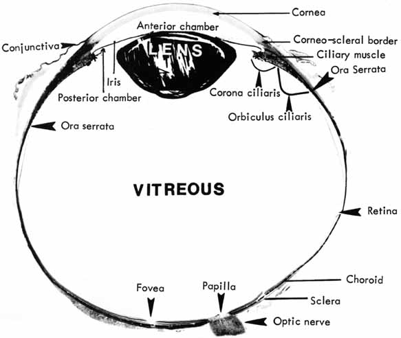
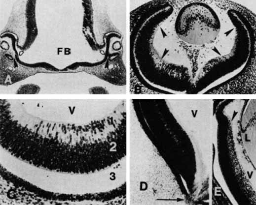
1. McDonnell JM: Ocular embryology and anatomy. In Ryan SJ, Ogden TE (eds): Retina, Vol 1. St. Louis: CV Mosby, 1989:11–15

2. Nussbaum JJ, Pruett RC, Delori FC: Historic perspectives—Macular yellow pigment: The first 200 years. Retina 1:296, 1981

3. Straatsma BR, Foos RY, Spencer LM: The retina—Topography and clinical correlations. In the New Orleans Academy of Ophthalmology Symposium on Retina and Retinal Surgery. St. Louis: CV Mosby, 1969

4. Straatsma BR, Landers MB, Kreiger AE: The ora serrata in the adult human eye. Arch Ophthalmol 80:3, 1968

5. White MH, Lambert HM, Kincaid MC, et al: The ora serrata and the spiral of Tillaux—Anatomic relationship and clinical correlation. Ophthalmology 96:608, 1989

6. Smiddy WE, Michels RG, Green WR: Lens and peripheral retinal relationships during vitrectomy. Retina 11:199, 1991

7. Hogan MJ: The vitreous, its structure, and relation to the ciliary body and retina. Invest Ophthalmol Vis Sci 2:418, 1963

8. Teng CC, Chi HH: Vitreous changes and the mechanism of retinal detachment. Am J Ophthalmol 44:335, 1957

9. Schepens CL: Vitreous changes in retinal detachment. In Schepens CL, Nutens A (eds): The Vitreous and Vitreoretinal Interface. New York: Springer-Verlag, 1987:85–107

10. Neubauer AS, Priglinger S, Ullrich S, et al: Comparison of foveal thickness measured with the retinal thickness analyzer 
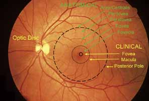
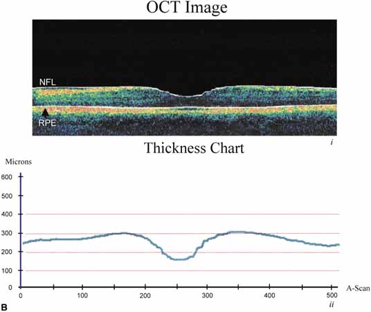
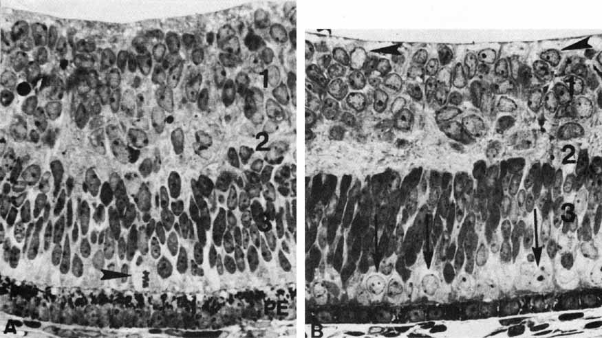
and optical coherence tomography. Retina 21:596–601, 2001

11. Blanks JC: Morphology of the retina. In Ryan SJ, Ogden TE (eds): Retina, Vol 1 St. Louis: CV Mosby, 1989:37–52

12. Osterberg G: Topography of the layer of rods and cones in the human retina. Acta Ophthalmol (suppl) 6:1, 1935

13. Yamada E: Some structural features of the fovea centralis in the human retina. Arch Ophthalmol 82:151, 1969

14. Hogan MJ, Alvarado JA, Weddell JE: Histology of the Human Eye—An Atlas and Textbook. Philadelphia: WB Saunders, 1971

15. Handelman GJ, Snodderly DM, Krinsky NI, et al: Biological control of primate macular pigment—Biochemical and densitometric studies. Invest Ophthalmol Vis Sci 32:257, 1991

16. Bone RA, Landrum JT, Fernandez L, Tarsis SL: Analysis of the macular pigment by HPLC—Retinal distribution and age study. Invest Ophthalmol Vis Sci 29:843, 1988

17. Snodderly DM, Handelman GJ, Adler AJ: Distribution of individual macular pigment carotenoids in central retina of macaque and squirrel monkeys. Invest Ophthalmol Vis Sci 32:268, 1991

18. Bone RA, Landrum JT, Mayne ST, et al: Macular pigment in donor eyes with and without AMD: A case-control study. Invest Ophthalmol Vis Sci 42:235–240, 2001

19. Thomson LR, Toyoda Y, Langner A, et al: Elevated retinal zeaxanthin and prevention of light-induced photoreceptor cell death in quail. Invest Ophthalmol Vis Sci 43:3538–3549, 2002

20. Bec P, Ravault M, Arne JL, Trepsat C: In the Fundus Periphery. New York: Masson Publishing, 1985:5

21. Byer NE: Cystic retinal tuft and miscellaneous peripheral retinal findings. In Jakobiec FA, Albert DM (eds): The Principles and Practice of Ophthalmology—Clinical Practice. Philadelphia: WB Saunders, 1993:1064–1070

22. Straatsma BR, Foos RY, Feman SS: Degenerative diseases of the peripheral retina. In Duane TD (ed): Clinical Ophthalmology, Vol 3, p 1. Hagerstown, MD:. Harper & Row, 1976

23. Spencer LM, Foos RY, Straatsma BR: Enclosed bays of the ora serrata—Relationship to retinal tears. Arch Ophthalmol 83:421, 1970

24. Spencer LM, Foos RY, Straatsma BR: Meridional folds, meridional complexes, and associated abnormalities of the peripheral retina. Am J Ophthalmol 70:697, 1970

25. Sebag J: Age-related changes in human vitreous structure. Graefes Arch Clin Exp Ophthalmol 225:89, 1987

26. Sebag J: Aging of the vitreous. Eye 1:254, 1987

27. Pei YF, Smelser GK: Some fine structural features of the ora serrata region in primate eyes. Invest Ophthalmol Vis Sci 7:672, 1968

28. Wise GN, Dollery CT, Henkind P: The Retinal Circulation. New York: Harper & Row, 1971

29. Duke-Elder S, Cook C: System of Ophthalmology, Vol III, Part I, Embryology. St. Louis: CV Mosby, 1963

30. Mann I: The Development of the Human Eye. New York: Grune & Stratton, 1964

31. Edward DP, Kaufman LM: Anatomy, development, and physiology of the visual system. Pediatr Clin N Am 50:1–23, 2003

32. Cepko CL, Austin CP, Yang X, et al: Cell fate determination in the vertebrate retina. Proc Natl Acad Sci USA 93:589–595, 1996

33. Lopashov CV, Stroeva OG: Development of the Eye—Experimental Studies. New York: Daniel Davey & Co, 1964

34. Vetter ML, Moore KB: Becoming glial in the neural retina. Developmental Dynamics 221:146–153, 2001

35. Hollenberg MJ, Spira AW: Early development of the human retina. Can J Ophthalmol 7:472, 1972

36. Spira AW, Hollenberg MJ: Human retinal development—Ultrastructure of the inner retinal layers. Dev Biol 31:1, 1973. Leventhal AG: Evidence that retinal ganglion cell density affects foveal development. Perspect Dev Neurobiol 3:203–211, 1996

37. Galli-Resta L: Putting neurons in the right places: local interactions in the genesis of retinal architecture. Trends in Neuroscience 25:638–643, 2002

38. Rapaport DH, Rakic P, Lavail MM: Spatiotemporal gradients of cell genesis in the primate retina. Prospectives on Developmental Neurobiology 3:147–159, 1996

39. Shatz CJ: Emergence of order in visual system development. Proc Natl Acad Sci USA 93:602–608, 1996

40. Wong RO, Chernjavsky A, Smith SJ, et al: Early functional neural networks in the developing retina. Nature 374:716–718, 1995

41. Nag TC, Wadhwa S: Expression of GABA in the fetal, postnatal, and adult human retina: an 
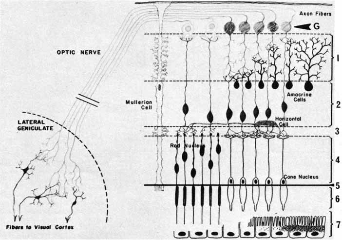
immunohistochemical study. Vis Neurosci 14:425–432, 1997

42. Catsicas M, Mobbs P: GABAb receptors regulate chick retinal calcium waves. J Neurosci 21:897–910, 2001

43. Leventhal AG: Evidence that retinal ganglion cell density affects foveal development. Perspect Dev Neurobiol 3:203–211, 1996

44. Hendrickson A, Kupfer C: The histogenesis of the fovea in the macaque monkey. Am J Med Sci 272:746, 1976

45. Peters A, Palay SL, Webster H deF: The Fine Structure of the Nervous System: The Neurons and Supporting Cells. Philadelphia: WB Saunders, 1976

46. Farquhar MG, Palade GE: Junctional complexes in various epithelia. J Cell Biol 17:375, 1963

47. Bennett MUL: A comparison of electrically mediated transmission. In Pappas GD, Purpura DP (eds): Structure and Function of Synapses. New York: Raven Press, 1972:221–256

48. Friend DS, Gilula NB: Variations in tight and gap junctions in mammalian tissues. J Cell Biol 53:758, 1972

49. Becker D, Bonness V, Mobbs P: Cell coupling in the retina: patterns and purpose. Cell Biology International 22:781–792, 1998

50. Dowling JE, Boycott BB: Organization of the primate retina—Electron microscopy. Proc R Soc Ser B 166:80, 1966

51. Raviola E, Gilula NB: Gap junctions between photoreceptor cells in the vertebrate retina. Proc Natl Acad Sci USA 70:1677, 1973

52. Kolb H, Famiglietti EV: Rod and cone pathways in the inner plexiform layer of cat retina. Science 186:47, 1974

53. Witkovsky P, Owen WG, Woodworth M: Gap junctions among the perikarya, dendrites, and axon terminals of the luminosity-type horizontal cell of the turtle retina. J Comp Neurol 216:359, 1983

54. Naka KI, Christensen BN: Direct electrical connections between transient amacrine cells in the catfish retina. Science 214:464, 1981

55. Cook JE, Becker DL: Gap junctions in the vertebrate retina. Microsc Res Tech 31:408–419, 1995

56. Witkovsky P, Stell WK: Retinal structure in the smooth dogfish Mustelus canis—Electron microscopy of serially sectioned bipolar cell synaptic terminals. J Comp Neurol 150:147, 1973

57. Famiglietti EV, Kolb H: A bistratified amacrine cell and synaptic circuitry in the inner plexiform layer of the retina. Brain Res 84:293, 1975

58. Massey SC, Mills SL: Gap junctions between AII amacrine cells and calbindin-positive bipolar cells in the rabbit retina. Vis Neurosci 16:1181–1189, 1999

59. Mills SL, Massey SC: A series of biotinylated tracers distinguishes three types of gap junctions in retina. J Neurosci 20:8629–8636, 2000

60. Iuvone PM: Neurotransmitters and neuromodulators in the retina--Regulation, interaction, and cellular effects. In Adler R, Farber D (eds): The Retina—A Model for Cell Biology Studies, Part II. Orlando: Academic Press, 1986:1–72

61. Weiler R, Ball AK, Ammermuller J: Neurotransmitter systems in the turtle retina. In Osborne N, Chader G (eds): Progress in Retinal Research, Vol 10. Oxford: Pergamon Press, 1991:1–26

62. Crooks J, Kolb H: Localization of GABA, glycine, glutamate, and tyrosine hydroxylase in the human retina. J Comp Neurol 315:287, 1992

63. Zucker CL, Ehinger B: Heterogeneity of receptor immunoreactivity at synapses of glycine-utilizing neurons. Proc R Soc Lond 249:89, 1992

64. Sun H, Crossland WJ: Quantitative assessment of localization and colocalization of glutamate, aspartate, glycine, and GABA immunoreactivity in the chick retina. Anat Rec 260:158–179, 2000

65. Johnson J, Rickman DW, Brecha NC: Somatostatin and somatostatin subtype 2A expression in the mammalian retina. Microsc Res Tech 50:103–111, 2000

66. Dowling JE: The Retina—An Approachable Part of the Brain. Cambridge: Harvard University Press, 1987

67. Gray EG: Electron microscopy of presynaptic organelles of the spinal cord. J Anat 97:101, 1963

68. Whittaker VP: The use of synaptosomes in the study of synaptic and neural membrane function. In Pappas GD, Purpura DP (eds): Structure and Function of Synapses. New York: Raven Press, 1972:87–100

69. Vollrath L, Spiwoks-Becker I: Plasticity of retinal ribbon synapses. Microsc Res Tech 35:472–487, 1996

70. Wagner HJ: Presynaptic bodies (“ribbons”): from ultrastructural observations to molecular perspectives. Cell Tissue Res 287:434–446, 1997

71. Yazulla S, Kleinschmidt J: Carrier-mediated release of GABA from retinal horizontal cells. Brain Res 263:63, 1983

72. Pfenninger KH: The cytochemistry of synaptic densities: II. Proteinaceous components and the mechanism of synaptic connectivity. J Ultrastruct Res 35:451, 1971

73. Fawcett DW: A Textbook of Histology, 11th ed. Philadelphia: WB Saunders, 1986

74. Kuffler SW, Nicholls JG: From Neuron to Brain—A Cellular Approach to the Function of the Nervous System. Sutherland: MA, Sinauer Associates Publishers, 1976

75. Morigiwa K, Vardi N: Differential expression of ionotropic glutamate receptor subunit in the outer retina. J Comp Neurol 405:173–184, 1999

76. Lin B, Martin PR, Solomon SG, et al: Distribution of glycine receptor subunits on primate retinal ganglion cells: a quantitative analysis. Eur J Neurosci 12:4155–4170, 2000

77. Koulen P, Sassoe-Pognetto M, Grunert U, et al: Selective clustering of GABA(A) and glycine receptors in the mammalian retina. J Neurosci 16:2127–2140, 1996

78. Grunert U: Distribution of GABA and glycine receptors on bipolar and ganglion cells in the mammalian retina. Microsc Res Tech 50:130–140, 2000

79. Pattnaik B, Jellali A, Sahel J, et al: GABAC receptors are localized with microtubule-associated protein 1B in mammalian cone photoreceptors. J Neurosci 20:6789–6796, 2000

80. Shields CR, Tran MN, Wong RO, et al: Distinct ionotropic GABA receptors mediate presynaptic and postsynaptic inhibition in retinal bipolar cells. J Neurosci 20:2673–2682, 2000

81. Keyser KT, MacNeil MA, Dmitrieva N, et al: Amacrine, ganglion and displaced amacrine cells in the rabbit retina express nicotinic acetylcholine receptors. Vis Neurosci 17:743–752, 2000

82. Wheeler-Schilling TH, Marquordt K, Kohler K, et al: Expression of purinergic receptors in bipolar cells of the rat retina. Brain Res Mol Brain Res 76:415–418, 2000

83. Nguyen-Legros J, Versaux-Botteri C, Vernier P: Dopamine receptor localization in the mammalian retina. Mol Neurobiol 19:181–204, 1999

84. Panda-Jonas S, Jonas JB, Jakobczyk-Zmija M: Retinal photoreceptor density decreases with age. Ophthalmology 102:1853–1859, 1995

85. Mohand-Said S, Hicks D, Leveillard T, et al: Rod-cone interactions: developmental and clinical significance. Progress in Retinal and Eye Res 20:451–467, 2001

86. Dunaief JL, Dentchev T, Ying G, et al: The role of apoptosis in age-related macular degeneration. Arch Ophthalmol 120:1435–1442, 2002

87. Missotten L: The ultrastructure of the human retina. Editions Arscia SA. Bruxelles: Uitgaven N V Presses academiques europeennes S C, 1965

88. Missotten L: L'ultrastructure des cones de la retine humaine. Bull Soc Belge Ophtalmol 132:472, 1963

89. Cohen AI: Rods and cones and the problem of visual excitation. In Straatsma BR, Hall MO, Allen RA, Crescitelli F (eds): The Retina—Morphology, Function and Clinical Characteristics. Berkeley, Los Angeles: University of California Press, 1969:31–62

90. Hargrave PA: Molecular dynamics of the rod cell. In Adler R, Farber D (eds): The Retina—A Model for Cell Biology Studies, Part I. Orlando: Academic Press, 1986:207–237

91. Wald G, Brown PK: Human rhodopsin. Science 127:222, 1958

92. Young RW: The renewal of photoreceptor cell outer segments. J Cell Biol 33:61, 1967

93. Hogan MJ, Wood I, Steinberg RH: Phagocytosis by pigment epithelium of human retinal cones. Nature 252:305, 1974

94. Besharse JC: Photosensitive membrane turnover in differentiated membrane domains and cell-cell interaction. In Adler R, Farber D (eds): The Retina—A Model for Cell Biology Studies, Part I. Orlando: Academic Press, 1986:297–352

95. Young RW: Visual cells and the concept of renewal. Invest Ophthalmol Vis Sci 15:700, 1976

96. Mullen RJ, LaVail MM: Inherited retinal dystrophy—Primary defect in pigment epithelium determined with experimental rat chimeras. Science 192:799, 1976

97. Anderson DH, Fisher SK: The photoreceptors of diurnal squirrels—Outer segment structure, disc shedding, and protein renewal. J Ultrastruct Res 55:119, 1976

98. Hogan MJ, Wood I, Steinberg RH: Phagocytosis by pigment epithelium of human retinal cones. Nature 252:305, 1974

99. Adler AJ, Severin KM: Proteins of the bovine interphotoreceptor matrix—Tissues of origin. Exp Eye Res 2:755, 1981

100. Hewitt AT, Adler R: The retinal pigment epithelium and interphotoreceptor matrix—Structure and specialized functions. In Ryan SJ, Ogden TE (eds): Retina, Vol 1. St. Louis: CV Mosby, 1989:57–64

101. Webster H deF, Ames A: Reversible and irreversible changes in the fine structure of nervous tissue during oxygen and glucose deprivation. J Cell Biol 26:885, 1965

102. Lessell S, Kuwabara T: Phosphatase histochemistry of the eye. Arch Ophthalmol 71:851, 1964

103. Matsusaka T: ATPase activity in the ciliary rootlet of the human retinal rods. J Cell Biol 33:203, 1967

104. Fine BS, Zimmerman LE: Observation on the rod and cone layer of the human retina—A light and electron microscopic study. Invest Ophthalmol Vis Sci 2:446, 1963

105. Uga S, Nakao F, Mimura M, Ikui H: Some new findings on the fine structure of the human photoreceptors cells. J Electron Microse (Tokyo) 19:71, 1970

106. Kuwabara T: Microtubules in the retina. In Rohen JW (ed): The Structure of the Eye, II Symp. Stuttgart: Schattauer-Verlay, 1965:69–84

107. Spitznas M: Zur Feinstruktur der sog. Membrana limitans externa der menschilichen Retina. Graefes Arch Clin Exp Ophthalmol 180:44, 1970

108. Kolb H, Famiglietti EV: Rod and cone pathways in retina of cat. Invest Ophthalmol Vis Sci 15:935, 1976

109. Apple DJ, Rabb MF: Ocular Pathology—Clinical Applications and Self-Assessment, 3rd ed. St. Louis: CV Mosby, 1985

110. Fine BS, Zimmerman LE: Müller's cells and the “middle limiting membrane” of the human retina—An electron microscopic study. Invest Ophthalmol Vis Sci 1:304, 1962

111. Boycott BB, Kolb H: The connections between bipolar cells and photoreceptors in the retina of the domestic cat. J Comp Neurol 148:91, 1973

112. Villegas GM: Ultrastructure of the human retina. J Anat 98:501, 1964

113. Kolb H, Linberg KA, Fisher SK: Neurons of the human retina—A Golgi study. J Comp Neurol 318:147, 1992

114. Mariani AP: Giant bistratified bipolar cells in the monkey retina. Anat Rec 206:215, 1983

115. Rodieck RW: The primate retina. Comp Primate Biol 4(Neurosciences) 203, 1988

116. Allen RA: The retinal bipolar cells and their synapses in the inner plexiform layer. In Straatsma BR, Hall MO, Allen RA, Crescitelli F (eds): The Retina--Morphology, Function and Clinical Characteristics. Berkeley, Los Angeles: University of California Press 1969:101–143

117. Fine BS: Synaptic lamellae in the human retina—An electron microscopic study. J Neuropathol Exp Neurol 22:255, 1963

118. Wu SM, Gao F, Maple BR: Integration and segregation of visual signals by bipolar cells in the tiger salamander retina. In Kolb H, Ripps H, Wu S (eds): Progress in Brain Research, Vol 131. Amsterdam: Elsevier Science BV, 2001:125–143

119. Kolb H: Organization of the outer plexiform layer of the primate retina: Electron microscopy of Golgi-impregnated cells. Philos Trans R Soc Lond (Biol) 258:261, 1970

120. Kolb H, Nelson R, Ahnelt P, et al: Cellular organization of the vertebrate retina. In Kolb H, Ripps H, Wu S (eds): Progress in Brain Research, Vol 131. Amsterdam: Elsevier Science BV, 2001:3–26

121. Boycott BB, Hopkins JM, Sperling HG: Cone connections of the horizontal cells of the rhesus monkey's retina. Proc R Soc Lond (Biol) 299:345, 1987

122. Yamada E: Some observations on the fine structure of the human retina. Fukuoka Acta Med 57:163, 1966

123. Kolmer W: Uber Krystalloide in Nervenzellen der menschlichen Netzhaut. Anat Anz 51:314, 1918

124. Yoshida M: The fine structure of the so-called crystalloid body of the human retina as observed with the electron microscope. J Electron Microsc (Tokyo) 14:285, 1966

125. Uga S, Ikui H: Some observations on the Kolmer's crystalloid of the human retina. J Electron Microsc (Tokyo) 18:153, 1969

126. Dietrich CE: Feinstructurelle Untersuchungen and den Horizontalzellen der menschlichen Netzhaut. Z Zellforsch 98:277, 1969

127. Yamada E, Ishikawa T: The fine structure of the horizontal cells in some vertebrate retinae. Cold Spring Harb Symp Quant Biol 30:383, 1965

128. Kolb H, Ahnelt P, Fisher SK, et al: Chromatic connectivity of the three horizontal cell types in the human retina. Invest Ophthalmol Vis Sci (suppl) 30:348, 1989

129. Dacey DM, Lee BB, Stafford DK, et al: Horizontal cells of the primate retina: cone specificity without spectral opponency. Science 271:656–659, 1996

130. Wassle H, Dacey DM, Haun T, et al: The mosaic of horizontal cells in the macaque monkey retina: with a comment on biplexiform ganglion cells. Vis Neurosci 17:591–608, 2000

131. Polyak SL: The Retina. Chicago: University of Chicago Press, 1941

132. Weiler R, He S, Vaney DI: Retinoic acid modulates gap junctional permeability between horizontal cells of the mammalian retina. Eur J Neurosci 11:3346–3350, 1999

133. Xin D, Bloomfield SA: Dark- and light-induced changes in coupling between horizontal cells in mammalian retina. J Comp Neurol 405:75–87, 1999

134. Dowling JE, Boycott BB: Neural connections of the retina: Fine structure of the inner plexiform layer. Cold Spring Harb Symp Quant Biol 30:393, 1965

135. Hughes A, Wieniawa-Narkiewicz E: A newly identified population of presumptive microneurons in the cat retinal 
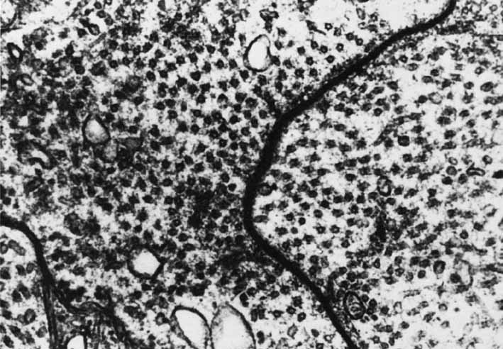
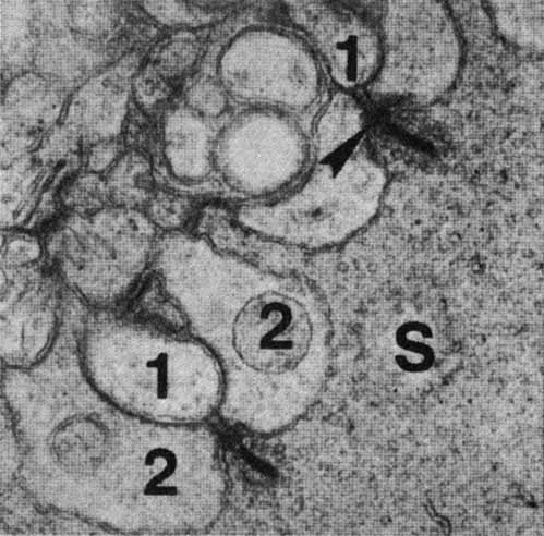
ganglion cell layer. Nature 284:468, 1980

136. Wong ROL, Hughes A: The morphology, number, and distribution of a large population of confirmed displaced amacrine cells in the adult cat retina. J Comp Neurol 255:159, 1987

137. Yoshida K, Watanabe D, Ishikane H, et al: A key role of starburst amacrine cells in originating retinal directional selectivity and optokinetic eye movement. Neuron 30:771–780, 2001

138. Zucker CL, Ehinger B: Synaptic connections involving immunoreactive glycine receptors in the turtle retina. Vis Neurosci 10:907, 1993

139. Gallego A: Horizontal and amacrine cells in the mammalian's retina. Vision Res (suppl) 3:33, 1971

140. Curio CA, Allen KA: Topography of ganglion cells in human retina. J Comp Neurol 300:5, 1990

141. Schein SJ: Anatomy of macaque fovea and spatial densities of neurons in foveal representation. J Comp Neurol 269:479, 1988

142. Kolb H, Marshak D: The midget pathways of the primate retina. Documenta Ophthalmologica 106:67–81, 2003

143. Polyak S: The vertebrate visual system. In Kluver H (ed): The Vertebrate Visual System. Chicago: University of Chicago Press, 1957

144. Bunt A, Minckler D: Displaced ganglion cells in the retina of the monkey. Invest Ophthalmol Vis Sci 16:95, 1977

145. Dowling JE, Boycott BB: Retinal ganglion cells: A correlation of anatomical and physiological approaches. In Straatsma BR, Hall MO, Allen RA, Crescitelli F (eds): The Retina--Morphology, Function and Clinical Characteristics. Berkeley, Los Angeles: University of California Press, 1969:145–161

146. Kenyon GT, Marshak DW: Gap junctions with amacrine cells provide a feedback pathway in ganglion cells within the retina. Proc R Soc Lond B Biol Sci 265:919–925, 1998

147. Wagner HG, MacNichol EFJr , Wolbarsht ML: The response properties of single ganglion cells in the goldfish retina. J Gen Physiol 43:45, 1960

148. Wiesel TN, Hubel DH: Spatial and chromatic intersections in the lateral geniculate body of the rhesus monkey. J Neurophysiol 29:1115, 1966

149. Gallego A: Connections transversales au niveau des couches plexiformes de la retine. Actual Neurophysiol 6:5, 1965

150. Brown JE, Major D: Cat retinal ganglion cell dendritic fields. Exp Neurol 15:70, 1966

151. Shapley R, Perry VH: Cat and monkey retinal ganglion cells and their visual functional roles. Trends Neurosci 9:229, 1986

152. Boycott GG, Wassle H: The morphological types of ganglion cells of the domestic cat's retina. J Physiol (Lond) 240:397, 1974

153. Berson E: Electrical phenomena in the retina. In Moses R, Hart W (eds): Adler's Physiology of the Eye. St. Louis: CV Mosby, 1987:506–567

154. Abe H, Iwata K: Checkerboard pattern reversal VER in the assessment of glaucomatous field defects. Acta Soc Ophthalmol Jpn 80:829, 1976

155. Quigley HA, Sanchez RM, Dunkelberger GR, et al: Chronic glaucoma selectively damages large optic nerve fibers. Invest Ophthalmol Vis Sci 28:913, 1987

156. Provencio I, Rodriguez IR, Jiang G, et al: A novel human opsin in the inner retina. J Neurosci 20:600–605, 2000

157. Hattar S, Liao HW, Takao M, et al: Melanopsin-containing retinal ganglion cells: architecture, projections, and intrinsic photosensitivity. Science 295:1065–1070, 2002

158. Barinaga M: How the brain's clock gets daily enlightenment. Science 295:955–957, 2002

159. Provencio I, Rollag MD, Castrucci AM: Photoreceptive net in the mammalian retina. Nature 415:493, 2002

160. Lucas RJ, Freedman MS, Munoz M, et al: Regulation of the mammalian pineal by non-rod, non-cone, ocular photoreceptors. Science 284:505–507, 1999

161. Harman A, Abrahams B, Moore S, et al: Neuronal density in the human retinal ganglion cell layer from 1 to 77 years. Anat Rec 260:124–131, 2000

162. Jonas JG, Dichtl A: Evaluation of the retinal nerve fiber layer. Surv Ophthalmol 40:369–378, 1996

163. Pollock SC, Miller NR: The retinal nerve fiber layer. Int Ophthalmol Clin 26:201, 1986

164. Vrabec F: The temporal raphe of the human retina. Am J Ophthalmol 62:926, 1966

165. Quigley HA, Addicks EM: Quantitative studies of the retinal nerve fiber layer defects. Arch Ophthalmol 100:807, 1982

166. Radius RL: Thickness of the retinal nerve fiber layer in primate eyes. Arch Ophthalmol 98:1625, 1980

167. Varma R, Skaf M, Barron E: Retinal nerve fiber layer thickness in normal human eyes. Ophthalmology 103:2114–2119, 1996

168. Takamoto T, Schwartz B: Differences by quadrant of retinal nerve fiber layer thickness in healthy 
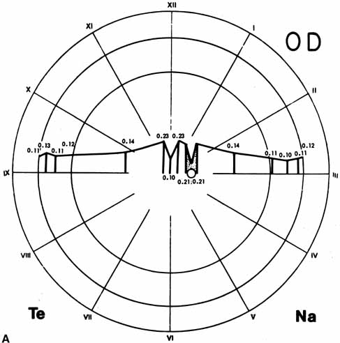
eyes. J Glaucoma 11:359–364, 2002

169. Bowd C, Weinreb RN, Williams JM, et al: The retinal nerve fiber layer thickness in ocular hypertensive, normal, and glaucomatous eyes with optical coherence tomography. Arch Ophthalmol 118:22–26, 2000

170. McLeod D, Marshall J, Kohner EM, et al: The role of axoplasmic transport in the pathogenesis of retinal cotton-wool spots. Br J Ophthalmol 61:177, 1977

171. Ochs S: Rate of fast axoplasmic transport in mammalian nerve fibers. J Physiol 227:627, 1972

172. Edstrom A, Mattsson H: Fast axonal transport in vitro in the sciatic system of the frog. J Neurochem 19:205, 1972

173. Guedes V, Schuman JS, Hertzmark E, et al: Optical coherence tomography measurement of macular and nerve fiber layer thickness in normal and glaucomatous human eyes. Ophthalmology 110:177–189, 2003

174. Tjon-Fo-Sang MJ, de Vries J, Lemij HG: Measurement by nerve fiber analyzer of retinal nerve fiber layer thickness in normal subjects and patients with ocular hypertension. Am J Ophthalmol 122:220–227, 1996

175. Aydin A, Wollstein G, Price LL, et al: Optical coherence tomography assessment of retinal nerve fiber layer thickness changes after glaucoma surgery. Ophthalmology 110:1506–1511, 2003

176. Uga S: Some structural features of the retina müllerian cells in the juxtaoptic nerve region. Exp Eye Res 19:105, 1974

177. Jean-Louis G, Katz BJ, Digre KB, et al: Acquired and progressive retinal nerve fiber layer myelination in an adolescent. Am J Ophthalmol 130:361–362, 2000

178. Rosen B, Barry C, Constable IJ: Progression of myelinated retinal nerve fibers. Am J Ophthalmol 127:4710473, 1999

179. Leys AM, Leys MJ, Hooymans JM, et al: Myelinated nerve fibers and retinal vascular abnormalities. Retina 16:89–96, 1996

180. Makarov FN, Hollender H, Stone J: The structure of interrelationships between neuroglial and ganglion cells in the retina. Neuroscience and Bahavioral Physiology 30:707–711, 2000

181. Boulder Committee: Embryonic vertebrate central nervous system. Revised terminology. Anat Rec 166:257, 1970

182. Weigert F: Beitrage zur Kenntnis der normalen menschlichen Neuroglia. Frankfurt am Main, Weisbrod, 1895

183. Palay SL, Chan-Palay V: Cerebellar Cortex, Cytology and Organization. New York: Springer-Verlag, 1974

184. Mori S, Leblond CP: Electron microscopic features and proliferation of astrocytes in the corpus callosum of the rat. J Comp Neurol 137:197, 1969

185. Vaughn JE, Hinds PL, Skoff RP: Electron microscopic studies of wallerian degeneration in rat optic nerves. I. The multipotential glia. J Comp Neurol 140:175, 1970

186. De Robertis D, Gerschenfeld HM: Submicroscopic morphology and function of glial cells. In Rev Neurobiol 3:1, 1961

187. Kuffler SW, Nicholls JG: The physiology of neuroglial cells. Ergeb Physiol 57:1, 1966

188. Palay SL: The role of neuroglia in the organization of the central nervous system. In Rodahl K, Issekutz B Jr (eds): Nerve as a Tissue. New York: Hoeber, 1966:3–10

189. Cammermeyer J: The life history of the microglial cell: A light microscopic study. In Ehrenpreis S, Solnitzky OC (eds): Neurosciences Research, Vol 3. New York: Academic Press, 1970:43–129

190. Matthews MA, Kruger L: Electron microscopy of nonneuronal cellular changes accompanying neural degeneration in thalamic nuclei of the rabbit. II. Reactive elements within the neuropil. J Comp Neurol 148:313, 1973

191. Stenwig AE: The origin of brain macrophages in traumatic lesions, wallerian degeneration, and retrograde degeneration. J Neuropathol Exp Neurol 31:696, 1972

192. Blakemore WF: Microglial reaction following thermal necrosis of the rat cortex: An electron microscope study. Acta Neuropathol 21:11, 1972

193. Inomata H: Electron microscopic observations on Müller's fiber in the human retina. Acta Soc Ophthalmol Jpn 69:2133, 1965

194. Hogan MJ, Feeney L: Ultrastructure of Müller cells and perivascular glia. Invest Ophthalmol Vis Sci 2:101a, 1963

195. Ogden TE: The glia of the retina. In Ryan WSJ, Ogden TE (eds): Retina, Vol I. St. Louis: CV Mosby, 1989:53–56

196. Uga S, Nakao F: The structure and function of the Müller cells in the human retina. II. The structure of the Müller cells in the normal retina. Acta Soc Ophthalmol Jpn 74:1018, 1970

197. Magalhaes MM: Functional cytoarchitecture of the retina Müller cell. In Yamada E, Mishima S (eds): The Structure of the Eye, III. Tokyo: Jpn J Ophthalmol, 1976:333–345

198. Magalhaes MM, Coimbra A: The rabbit retina Müller cell. A fine structural and cytochemical study. J Ultrastruct Res 39:310, 1972

199. Edwards RB, Adler AJ, Dev S, Claycomb RC: Synthesis of retinoic acid from retinol by cultured rabbit Müller cells. Exp Eye Res 54:481, 1992

200. Das SR, Bhardwaj N, Kjeldbye H, Gouras P: Müller cells of chicken retina synthesize 11-cis-retinol. Biochem J 285:907, 1992

201. Eisenfeld AJ, Bunt-Milam AH, Saari JC: Localization of retinoid-binding proteins in developing rat retina. Exp Eye Res 41:299, 1985

202. Milam AH, De Leeur AM, Gaur VP, Saari JC: Immunolocalization of cellular retinoic acid-binding protein to Müller cells and/or a subpopulation of GABA-positive amacrine cells. J Comp Neurol 296:123, 1990

203. Inomata H: Fine structure of cystoid degeneration in the peripheral retina. Acta Soc Ophthalmol Jpn 69:1357, 1965

204. Gass JD: Muller cell cone, an overlooked part of the anatomy of the fovea centralis: hypotheses 
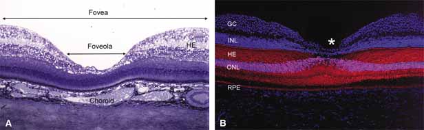
concerning its role in the pathogenesis of macular hole and foveomacular retinoschisis. Arch Ophthalmol 117:821–823, 1999

205. Hogan MJ, Feeney L: The ultrastructure of the retinal vessels. III. Vascular-glial relationships. J Ultrastruct Res 9:47, 1963

206. Wolter JR: The cells of Remak and the astroglia of the normal human retina. Arch Ophthalmol 53:832, 1955

207. Wolter JR: Glia of the human retina. Am J Ophthalmol 48:370, 1959

208. Wolter JR: The astroglia of the human retina and other glial elements of the retina under normal and pathologic conditions. Am J Ophthalmol 40:88, 1955

209. Provis JM: Development of the primate retinal vasculature. Progress in Retinal and Eye Research 20:799–821, 2001

210. Ramirez JM, Ramirez AI, Salazar JJ, et al: Changes of astrocytes in retinal ageing and age-related macular degeneration. Exp Eye Res 73:601–615, 2001

211. Vrabec F: Microglia in the monkey and rabbit retina. J Neuropathol Exp Neurol 29:217, 1970

212. Provis JM, Diaz CM, Penfold PL: Microglia in human retina: a heterogeneous population with distinct ontogenies. Perspect Dev Neurobiol 3:213–222, 1996

213. Roque RS, Rosales AA, Jingjing L, et al: Retina-derived microglial cells induce photoreceptor cell death in vitro. Brain Res 836:110–119, 1999

214. Smelser GK, Ishikawa T, Pei YF: Electron microscopic studies of intra-retinal spaces—Diffusion of particulate materials. In Rohen JW (ed): Structure of the Eye, II Symp. Stuttgart: Schattauer-Verlay, 1965:109–121

215. Iwasaki M, Myers KM, Rayborn ME, Hollyfield JG: Interphotoreceptor matrix in the human retina: Cone-like domains surround a small population of rod photoreceptors. J Comp Neurol 319:277, 1992

216. Hageman GS, Johnson LV: Structure, composition and function of the retinal interphotoreceptor matrix. In Osborne N, Chader G (eds): Progress in Retinal Research, Vol 10. Oxford: Pergamon Press, 1991:207–250

217. Jerdan JA, Kao L, Glaser BM: The inner limiting membrane: A modified basement membrane? Invest Ophthalmol Vis Sci (suppl) 27:230a, 1986

218. Kohno T, Sorgente N, Ishibashi T, et al: Immunofluorescent studies of fibronectin and laminin in the human eye. Invest Ophthalmol Vis Sci 28:506, 1987

219. Foos RY: Vitreoretinal juncture: Topographical variations. Invest Ophthalmol Vis Sci 11:801, 1972

220. Anderson DR: Ultrastructure of the optic nerve head. Arch Ophthalmol 83:63, 1970

221. Bargmann W: Histologie und mikroskopische Anatomie des Menschen, 6th ed. Stuttgart: Georg Thieme Verlag, 1967

222. Archer D, Krill AE, Newell FW: Fluorescein studies of normal choroidal circulation. Am J Ophthalmol 69:543, 1970

223. Ashton N: The mode of development of the retinal vessels in man. In Cant JS (ed): The William MacKenzie Symposium on the Ocular Circulation in Health and Disease. St. Louis: CV Mosby, 1969:7–18

224. Cogan DG: Development and senescence of the human retinal vasculature. Trans Ophthalmol Soc UK 83:465, 1963

225. Mintz Hittner HA, Knight Nanan DM, Satriano DR, et al: A small foveal avascular zone may be an historic mark of prematurity. Ophthalmology 106:1409–1413, 1999

226. Mutlu F, Leopold IH: The structure of the human retinal vascular system. Arch Ophthalmol 71:93, 1964

227. Kuwabara T, Cogan DG: Studies of retinal vascular patterns. Part I. Normal architecture. Arch Ophthalmol 64:904, 1960

228. Justice J, Lehmann RP: Cilioretinal arteries. Arch Ophthalmol 94:1355, 1976

229. Maharajan VS, Dua HS, Scott R, et al: Unexpected visual improvement with a pinhole in central retinal artery occlusion and cilioretinal artery sparing fovea. Eye 16:194–196, 2002

230. Ernest JT: Macrocirculation and microcirculation of the retina. In Ryan SJ, Ogden TE (eds): Retina, Vol I. St. Louis: CV Mosby, 1989:65–66

231. Harris A, Cuilla TA, Chung HS, et al: Regulation of retinal and optic nerve blood flow. Arch Ophthalmol 116:1491–1495, 1998

232. Laties AM: Central retinal artery innervation: Absence of adrenergic innervation to the intra-ocular branches. Arch Ophthalmol 77:405, 1967

233. Laties AM: Neurovascular relationships in the retina. Anat Rec 163:216, 1969

234. Forster BA, Ferrari-Dileo G, Anderson DR: Adrenergic alpha 1 and alpha 2 binding sites are present in bovine retinal blood vessels. Invest Ophthalmol Vis Sci 28:1741, 1987

235. Denis P, Elena PP: Recepteurs beta-adrenergiques vasculares retiniens chez I'homme. Ophthalmologie 3:62, 1989

236. Ferrari-Dileo G, Davis EB, Anderson DR: Response of retinal vasculature to phenylephrine. Invest Ophthalmol Vis Sci 31:1181, 1990

237. Shin DH, Tsai CS, Parrow KA, et al: Vasoconstrictive effect of topical timolol on human retinal arteries. Graefes Arch Clin Exp Ophthalmol 229:298, 1991

238. Hogan MJ, Feeney L: Ultrastructure of the retinal vessels. Part I. The larger vessels. J Ultrastruct Res 9:10, 1963

239. Shakib M, Cunha-Vaz JG: Studies on the permeability of the blood-retinal barrier. IV. Junctional complexes of the retinal vessels and their role in the permeability of the blood-retinal barrier. Exp Eye Res 5:229, 1966

240. Gardner TW, Antonetti DA, Barber AJ, et al: The molecular structure and function of the inner blood-retinal barrier. Documenta Ophthalmologica 97:229–237, 1999

241. Ashton N: Studies on the retinal capillaries in relation to diabetic and other retinopathies. Br J Ophthalmol 47:521, 1963

242. Hogan MJ, Feeney L: The ultrastructure of the retinal blood vessels. II. The small vessels. J Ultrastruct Res 9:29, 1963

243. Green WR, Chan CC, Hutchins GM, Terry JM: Central retinal vein occlusion: A prospective histopathologic study of 29 eyes in 28 cases. Retina 1:27, 1981

244. Feist RM, Ticho BH, Shapiro MJ, Farber M: Branch retinal vein occlusion and quadrant variation in arteriovenous crossings. Am J Ophthalmol 113:664, 1992

245. Wong TY, Klein R, Nieto FJ, et al: Retinal microvascular abnormalities and 10-year cardiovascular mortality—a population-based case-control study. Ophthalmology 110:933–940, 2003

246. Weinberg DV, Egan KM, Seddon JM: Asymmetric distribution of arteriovenous crossings in the normal retina. Ophthalmology 100:31, 1993

247. Opremcak EM, Bruce RA: Surgical decompression of branch retinal vein occlusion via arteriovenous crossing sheathotomy: a prospective review of 15 cases. Retina 19:1–5, 1999

248. Zhao J, Sastry SM, Sperduto RD, et al: The Eye Disease Case-Control Study Group: Arteriovenous crossing patterns in branch retinal vein occlusion. Ophthalmology 100:423, 1993

249. Rassam SM, Patel V, Chen HC, et al: Regional retinal blood flow and vascular autoregulation. Eye 10:331–337, 1996

250. Rawji MH, Flanagan JG: Intraocular and interocular symmetry in normal retinal capillary perfusion. J Glaucoma 10:4–12, 2001

251. Toussaint D, Kuwabara T, Cogan DG: Retinal vascular patterns. II. Human retinal vessels studied in three dimensions. Arch Ophthalmol 65:575, 1961

252. Marquardt R: Contribution to topography and anatomy of the retinal vessels of the human 
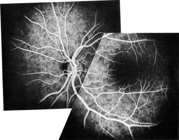
eye. Klin Monatsbl Augenheilkd 148:50, 1966

253. Michaelson IC: Retinal Circulation in Man and Animals. Springfield, IL:. Charles C Thomas, 1954

254. Duke-Elder S, Wybar KC: System of Ophthalmology, Vol II. The Anatomy of the Visual System. St. Louis: CV Mosby, 1961

255. Snodderly DM, Weinhaus RS, Choi JC: Neural-vascular relationships in central retina of macaque monkeys (Macaca fascicularis). J Neurosci 12:1169, 1992

256. Iwasaki M, Inomata H: Relation between superficial capillaries and foveal structures in the human retina. Invest Ophthalmol Vis Sci 27:1698, 1986

257. Henkind P: New observations on the radial peripapillary capillaries. Invest Ophthalmol 6:103, 1967

258. Henkind P: Radial peripapillary capillaries: Past-present-future. In Shimizu K (ed): Fluorescein Angiography. Tokyo: Igaku Shoin, 1974:91–95

259. Kornzweig AL, Elasoph I, Feldstein M: Selective atrophy of the radial peripapillary capillaries in chronic glaucoma. Arch Ophthalmol 80:696, 1968

260. Kuwabara T, Cogan DG: Retinal vascular patterns, Part VI. Mural cells of the retinal capillaries. Arch Ophthalmol 69:492, 1963

261. Martin AR, Bailie JR, Robson T, et al: Retinal pericytes control expression of nitric oxide synthase and endothelin–1 in microvascular endothelial cells. Microvasc Res 59:131–139, 2000

262. Bandopadhyay R, Orte C, Lawrenson JG, et al: Contractile proteins in pericytes at the blood-brain and blood-retinal barriers. J Neurocytology 30:35–44, 2001

263. Chen Q, Anderson DR: Effect of CO2 on intracellular pH and contraction of retinal capillary pericytes. Invest Ophthalmol Vis Sci 38:643–651, 1997

264. Cogan DG, Toussaint D, Kuwabara T: Retinal vascular patterns, Part IV. Diabetic retinopathy. Arch Ophthalmol 66:366, 1961

265. Hammes H, Lin J, Renner O, et al: Pericytes and the pathogenesis of diabetic retinopathy. Diabetes 51:3107–3112, 2002

266. Circulating anitpericyte autoantibodies in diabetic retinopathy. Retina 19:390–400, 1999

267. Leuenberger PM: Ultrastructure of the aging retinal vascular system with special reference to quantitative and qualitative changes of capillary basement membranes. Gerontologia 19:1, 1973

Back to Top