|
|
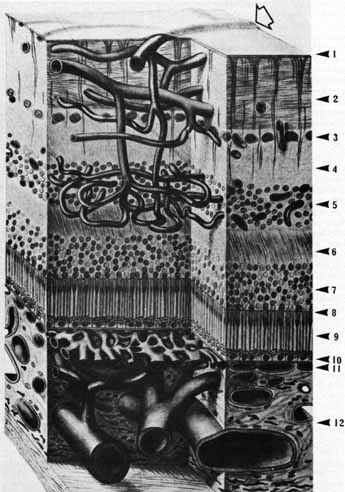
| Fig. 44 Podesta's scheme of the retinal vasculature demonstrates a speculative bilevel capillary arrangement, with an even-depth distribution of postarteriolar and prevenular capillaries. There is no vasculature between the inner nuclear layer and the choriocapillaris. Retinal vein shows how the major vessels lie immediately beneath and elevate the internal limiting membrane (open arrow). 1, inner limiting membrane; 2, nerve fiber layer; 3, ganglionic layer; 4, inner plexiform layer; 5, inner nuclear layer; 6, outer plexiform layer; 7, outer nuclear layer; 8, external limiting membrane; 9, layer of photoreceptor inner and outer segments; 10, pigmented epithelium; 11, choriocapillaris; 12, choroid. (Bargmann W: Histologie und mikroskopische Anatomie des Menschen, 6th ed. Stuttgart: Georg Thieme Verlag, 1967) |