|
|
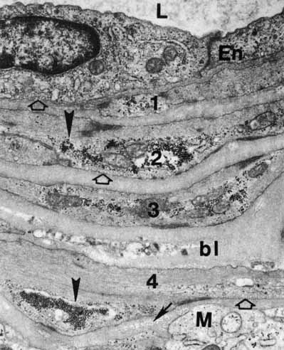
| Fig. 46 Part of an arterial will in the nerve fiber layer of the central retina with four layers of smooth muscle cells (1, 2, 3, 4) embedded in thick basal laminalike substance (bl) En, endothelial cell; L, lumen. Glycogen forms masses in the muscle cells (arrowheads) and small clusters in the Müller cell (M) processes around the vessel. Collagen fibrils (arrow) are in the basal lamina lining the Müller cell. Pinocytotic vesicles are on the abluminal surfaces of the muscle cells, as well as in the endothelial cytoplasm (open arrows) (original magnification ×24,000). |