|
|
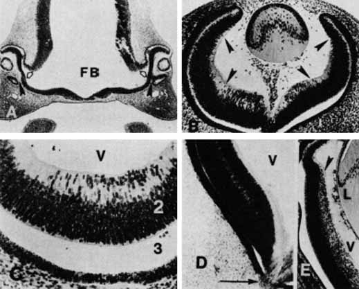
| Fig. 17 A. At 33 days' gestation, embryonic retina is separated from the future pigmented epithelium by the space of the primary optic vesicle (asterisk), which communicates with the cavity of the forebrain (FB) by means of the optic stalks (open arrows). The embryonic or fetal fissure (arrowheads) provides the site of entrance of blood vessels into the optic cup (original magnification ×60). B. At 39 days' gestation, the hyaloid vessels extend from the embryonic fissure through the vitreous cavity to reach the lens. The posterior retina shows differentiation into the neuroepithelium with two distinct neuroblastic layers. The peripheral retina (arrowheads) continues as a monolayer and has not yet formed the bilayered neuroepithelium (original magnification ×150). C. At 35 days' gestation, the retina has the beginning of an inner nuclear zone, the inner neuroblastic layer (1). The outer neuroblastic layer (2) continues to show cell division. The neuroepithelium remains unattached to the underlying multilayered pigmented epithelium. The wide space of the site of the primary optic vesicle (3) is caused by fixation and tissue preparation artifact. The internal limiting membrane is formed and separates the neuroepithelium from the vitreous (V) (original magnification ×300). D. At 41 days' gestation, the neuroepithelium has distinct outer and inner neuroblastic layers. The axons of the ganglion cells have begun to form the nerve fiber layer as they migrate toward the embryonic fissure, where they will form the optic nerve (arrow). V, vitreous body (original magnification ×200). E. At 10 weeks' gestation (54 mm), the division into the two neuroblastic layers has extended peripherally up to the future ora serrata (arrowhead). Retinal maturation progresses from the posterior pole to the anterior region of the optic cup and from the inner ganglion cell layer outwardly to the photoreceptors. L, lens; V, vitreous body (original magnification ×100). |