|
|
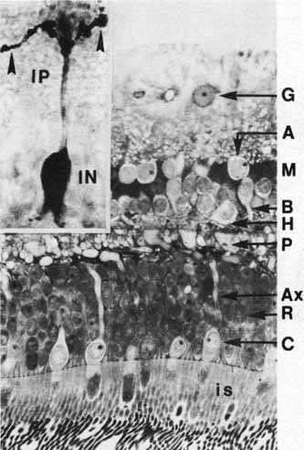
| Fig. 26 Thin section of the retina showing location of its neural elements. A, amacrine cell nucleus; B, bipolar cell nucleus; C, cone nucleus; Ax, cone axon; G, ganglion cell nucleus; H, horizontal cell nucleus; M, Müller cell nucleus; P, cone pedicles; R, rod nucleus; is, photoreceptor inner segments (original magnification ×720). Inset. Golgi-impregnated rod bipolar cell. Arrowheads indicate telodendritic expansions in the inner plexiform layer (IP). The nucleus is situated in the inner nuclear layer (IN) (original magnification ×1120). |