|
|
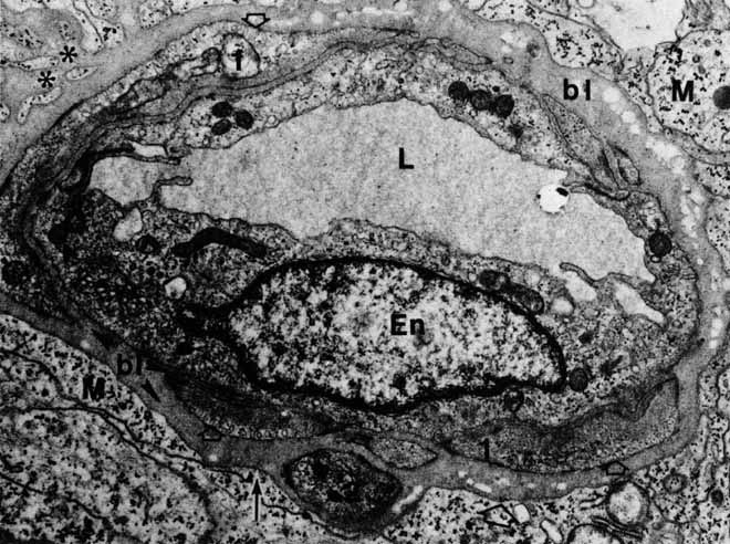
| Fig. 47 Postcapillary venule in the nerve fiber layer, with two interrupted layers of muscle cells (1, 2) embedded in basal lamina (bl). Müller cell (M) cytoplasm is fused to basal lamina (arrow), which also extends into the glial interstitial spaces (asterisks). Numerous pinocytotic vesicles line the abluminal surfaces of muscle cells (open arrows). En, endothelial cell nucleus; L, lumen (original magnification ×6000). |