|
|
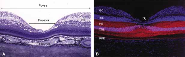
| Fig. 7 A. Light micrograph of human anatomic fovea and foveola stained with Richardson's methylene blue/azure II mixture. 10× objective. B. Immunofluorescence labeling of human cones (red with monoclonal antibody against cone-specific enolase) and their nuclei (pink) in the fovea. Note the tightly packed cones with long, thin outer segments and axons forming the fiber layer of Henle (HE). Other cell nuclei in the outer nuclear layer (ONL), inner nuclear layer(INL) and ganglion cell layer (GC) are stained blue with DAPI. Retinal pigment epithelium (RPE) shows auto fluorescence. Foveal pit (*) 10× objective. (Courtesy of Scheie Eye Institute) |