|
|
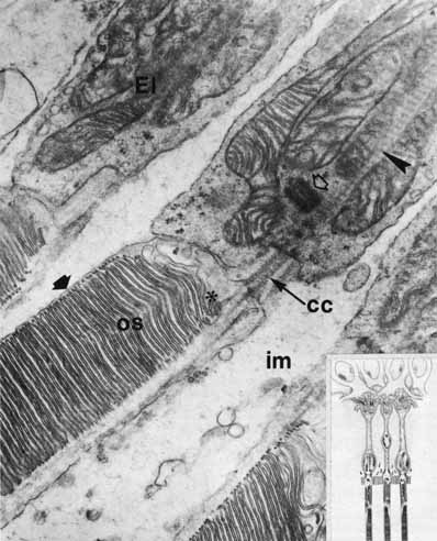
| Fig. 21 Electron micrograph of portions of rod inner and outer segments: connecting cilium (cc) between rod outer segment (os) and ellipsoid (El); basal body (centriole; open arrow); striated ciliary rootlets (arrowhead) between long mitochondria of inner segment; mucopolysaccharide–protein-containing matrix (im). Note rod saccules (discs) covered by plasma membrane (solid arrow). Asterisk indicates basal rod discs in formation (original magnification ×24,000). Inset. Schematic drawing of retinal receptors and interreceptor contacts. Rod spherule is in the middle. (Sjöstrand FS: Electronmicroscopy of the retina. In Smelser GK (ed): The Structure of the Eye. New York: Academic Press, 1961:1–28) |