|
|
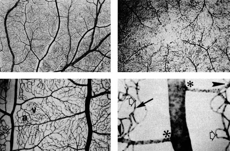
| Fig. 48 Blood vessels of the retina. Top left. Trypsin digest preparation of the human retinal vasculature. The darker, narrower channels surrounded by capillary-free-zones are arteries. Top right. Capillary bed around the macula. The second layer of capillaries is represented by the broken profiles. There is a capillary-free zone in the center. Bottom left. Equatorial zone of the retina. The artery is on the left, with a capillary-free zone. The vein is at the right. Note the interdigitations of the arteriolae efferens (a) with the venule efferens (v). Bottom right. An artery gives off arterioles (asterisks), which in turn give off capillaries (arrowheads). Endothelial cell nuclei are indicated by arrows; the bulging pericyte nuclei, by open arrows. (Courtesy of Dr. Paul Henkind) |